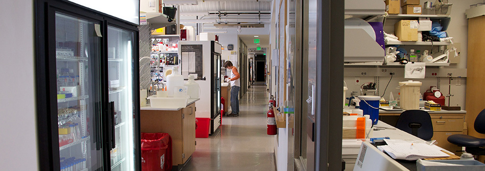
The Anatomy and Cell Biology Department has a collection of commonly used equipment. All of this equipment requires training prior to being used. If you wish to use any of this equipment please email or see Dennis Dunnwald (dennis-dunnwald@uiowa.edu), Room 1-339.
Sign up at (BookItLab)
Microscopes
- Nikon Inverted –1-251 Inside darkroom turn sliding door to the right 1/4 turn (Caretaker Dennis Dunnwald). This microscope is ideal for fluorescent of tissue culture on plastic plates.
- Nikon Inverted–1-418 (Caretaker Chuck Yeaman, located in tissue culture room). This microscope is ideal for fluorescent of tissue culture on plastic plates.
- Nikon Upright-1-370 (Caretaker Dennis Dunnwald). This microscope is ideal bright field imaging of slides. Equipped with a DS-Fi3 camera for taking epifluorescence GFP/RFP/DAPI images, bright field and color (such as HE staining) images. Suitable for slides only.
- Leica Thuder 3D Assay Microscope–1-558 (Caretaker Dennis Dunnwald.)
- Zeiss 700 Laser Scanning Confocal Microscope –1-560 (Caretaker Dennis Dunnwald and training.) Has 2 PMT, 4 laser lines, temperature and CO2 controls. Good for live and fixed imaging. Please Read: Zeiss Policy and
 Zeiss 700 User Guide
 - Zeiss Training Video Tutorials for 880 and 980 confocal microscopes
- Zeiss 880 Laser Scanning Confocal Microscope with Airyscan -1-249 (Caretaker Dennis Dunnwald. Training by Tom Moninger in the CMRF required). Has Airyscan (fast and super resolution scanning ability), 4 PMT, and 6 laser lines (allows FRET imaging), 405-pulsed laser for bleaching and ablation, temperature controls, several oil and water objectives for high-resolution imaging. Good for live and fixed imaging.
 Zeiss 880 User Guide
 - Zeiss 980 Laser Scanning Confocal Microscope with Airyscan 2 - 1-341 (Caretaker Dennis Dunnwald and training) Has Airyscan2 (fast and super resolution scanning ability), 4 PMT, and 6 laser lines (allows FRET imaging), 405-pulsed laser for bleaching and ablation, temperature controls, several oil and water objectives for high-resolution imaging. Good for live and fixed imaging.
 Zeiss 980 User Guide
 - Metamorph Software-dedicated Computer-1-251 Second Door (Caretaker Dennis Dunnwald. Engelhardt Lab is a heavy user). The morphometric analysis packages are useful in quantitatively evaluating fluorescent images. The system is not extremely user friendly and takes time to learn, but has useful cell scoring features. An alternative to this system is Fiji (NIH Image, which is free to download onto your computer).
Other equipment (Caretaker Dennis Dunnwald):
Sign up at BookItLab
- Licor Odyssey Imaging System –1-370 (IR Dye Westerns)
- Amersham™ Imager 600 series –1-370 Scanning Chemiluminescence Westerns
- Ultracentrifuge –1-400C Near Rob Cornell's Lab
- BioRad CFX96 Real-Time PCR machine –1-370
- Nanodrop Spectrophotometer (DNA/RNA Quantitation) – 1-440A
- Bacterial Shaker –1-370
- Autoclaves –1-503
- Washers and Drying Oven –1-503
- Ice Machine –1-503