Our videographer has produced programs for the AAO COVE program and the AAO Annual Video Program, N.E.I.E., N.J.M., Numerous in-house programs have also been produced for resident, fellow, nurse, and technician education. Examples may be viewed from the EyeRounds web site.
The Braley Auditorium is equipped with a dedicated computer for Internet and Power Point media. A closed circuit video system allows transparencies and opaque materials to be displayed, ophthalmic pathology microscope slides to be seen, videotapes shown, all through a multi-sync LCD large venue projector onto a 10' diagonal screen. This CCTV system is used during daily morning conference, weekly fluourescein and pathology conferences, and for monthly clinical conferences. Any lecture or presentation can be recorded for later viewing using Panopto software that uses screen capture for computer content, video of the presenter, and audio. Access to these lectures is for Iowa Residents, Fellows, Faculty and Alumni.
The Retina service also uses a video indirect system to clinically document pathology and for teaching. The system may also be taken into the OR/ASC and used during surgery.
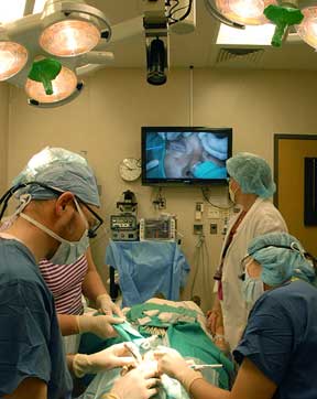
 Image right: Use of High definition Video in the Ophthalmic Procedure Suite
The Minor Procedure rooms here are equipped with movable arm mounted video cameras to record Oculoplastic procedures for teaching. Recently we installed a High Definition video system which yields outstanding image resolution.
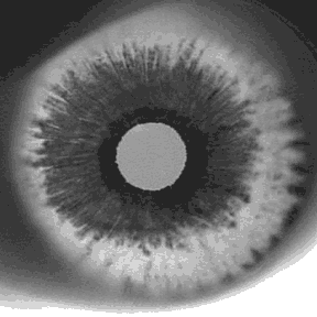
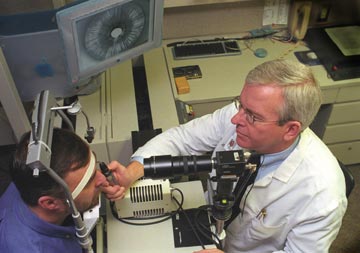
A unique instrument we utilize here is the Iowa Infrared Pupilometer that allows a clinician to view a patient's eyes in the dark through the use of infrared LEDs and an infrared sensitive video camera. Prisms allow a view of just a patient’s two eyes side by side. This device can also be used for transillumination of the iris and sclera to look at PDS and other iris abnormalities, iris cysts and tumors, scleral tumor location, foreign body location, Diabetes, Adie’s pupil, Pseudoexfoliation, and the Meibomian glands.


More about Transillumination
The Pediatric department uses video as a children's fixation device. Eight exam rooms are equipped with color monitors that are fed cartoons via a video/audio distribution amplifier. A footswitch instantly turns the cartoons with sound on and off. This cartoon fixation system has proven very helpful in maintaining a child's fixation during examination.
Some great examples of the video imaging performed at The University of Iowa is at gonioscopy.org.