- An intimal tear allows blood to enter the arterial wall and separate the intimal layer of the artery from the media, forming a hematoma
- Most commonly occur in the internal carotid artery 2-3 cm away from the bifurcation of the common carotid
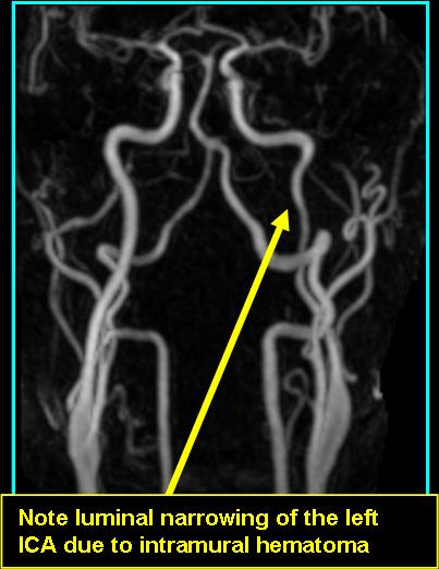
- Will show both luminal narrowing and aneurysmal dilatation on imaging
- On CT: contast will show double lumen, luminal narrowing, flap formation, and/or aneurysmal dilatation
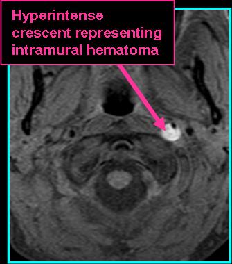
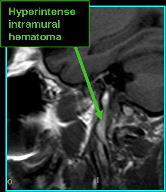
- On MR:
- T1 with fat-suppression will show intramural hematoma as a crescent
- Hematoma also visualized on T2
- MRA shows vessel tapering and aneurysmal dilatation
- Most commonly occurs spontaneously, etiology unknown
- May also occur in setting of trauma, although vertebral dissection is more common
- Some with familial connective tissue diseases may be predisposed
![]()
![]()
References
Hakimi R, Sivakumar S. Imaging of Carotid Dissection. Curr Pain Headache Rep. 2019;23(1):2. Published 2019 Jan 19. doi:10.1007/s11916-019-0741-9
Schievink WI. Spontaneous dissection of the carotid and vertebral arteries. N Engl J Med. 2001;344(12):898-906. doi:10.1056/NEJM200103223441206