General Considerations
-
Selective upper airway stimulation (UAS) using an implanted hypoglossal nerve stimulator has become a widely available surgical management for obstructive sleep apnea recalcitrant to continuous positive airway pressure (CPAP) treatment or for those patients for whom CPAP is not an option.
-
Clinical trials have shown that in carefully selected adult patients there are dramatic clinical improvements. One study showed a 68% decrease in the apnea-hypopnea index (AHI) and 70% decrease in oxygen desaturation index (Heiser et al. 2017).
-
Another study showed that UAS lead to significantly improved sleepiness, quality of life, and respiratory outcomes at 5 years (Woodson et al. 2018).
-
The device is only currently indicated for adults, but trials in children are ongoing (Diercks et al. 2018).
Anatomy
-
An intimate understanding of neck anatomy is required along with knowledge of branching patterns of the hypoglossal nerve and innervation of tongue musculature.
-
The goal is to include branches of the hypoglossal nerve that innervate muscles that protrude the tongue while excluding branches that retract the tongue (Heiser et al. 2017).
-
Branches to include:
-
Transverse branch (intrinsic muscle)
-
Vertical branch (intrinsic muscle)
-
Genioglossus oblique (extrinsic muscle)
-
Genioglossus horizontal (extrinsic muscle)
-
First cervical branch to geniohyoid (C1, extrinsic muscle)
-
-
Branches to exclude:
-
Styloglossus
-
Hyoglossus
-
Preoperative Preparations
-
Sleep study with AHI and oxygen desaturation index is required to assess sleep apnea and obstructive component.
-
Drug induced sleep endoscopy is required to assess dynamic airway collapse.
Nursing Considerations
-
Standard Oto Major tray
-
Oto micro tray
-
Vessel loops
-
Malleable retractors (thinnest ~ 1 cm retractor is required). Large Rich retractor needed for sense lead placement.
-
Operating microscope is required for hypoglossal nerve dissection.
-
Medtronic NIM nerve monitoring system is required for monitoring of hypoglossal nerve.
-
Medtronic tunneler, MDT Catheter passer (single use), 38 cm, ref 8591-38
-
Equipment required "off the field":
-
Bipolar stimulation electrodes x 2 (Xomed 8227304 and 8225401)
-
Debakey forceps to grasp and insert the electrodes
-
4x4 gauze to grasp the tongue
-
Marker
-
Local anesthetic
-
-
Prep and drape
-
Arms tucked
-
Shoulder roll
-
No foley catheter
-
Head turned to the right, prep from lower lip, down right neck and to xiphoid process
-
1010 sticky drape
-
Standard Ioban drape
-
Oto split drape
-
-
A sterile sleeve is required for the telemetry unit on to the field to test the device.
Anesthesia Considerations
-
Standard orotracheal intubation is used, ETT size is not important.
-
ETT taped to corner of left mouth.
-
No long acting paralytics should be used but short acting muscle relaxant is fine.
-
Preoperative antibiotic (ancef) is given.
Operative Technique
-
No monopolar cautery is used
-
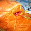
Patient preparation (see photos above):
-
Plan neck incision (stimulation electrode): 4-6 cm in length, 1 cm from midline, 2 cm below mandible
-
Plan implantable pulse generator (IPG) pocket: 5 cm inferior to clavicle, 5.5 cm in length
-
Plan chest incision (sensor electrode): inferior margin of right pectoralis major, 5th rib +/-1 intercostal space, 4-6 cm in length
-
Place NIM electrodes in tongue: grip tongue with gauze and use debakey forceps to place red electrodes in hyoglossus and blue electrodue in genioglossus
-
Place clear 1010 drape from along mandible up over mouth so that tongue can be visualized intraoperatively but is separate from the sterile field
-
Prep patient from lower lip to xyphoid
-
Place sterile towels and ioban drape
-
Place U drape
-
-
Neck incision
-
Divide skin and platysma, small inferior subplatysma flap
-
Identify and retract digastric tendon and submandibular gland
-
Identify mylohyoid and divide ranine vein as necessary
-
-
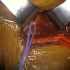
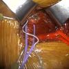
Hypoglossal nerve identification: place vessel loop to isolate nerve.
-
Hypoglossal nerve dissection and isolation:
-
Bring in microscope
-
Identify functional breakpoint of hypoglossal nerve using bipolar stimulator
-
Stimulator lead placement around inclusion branches of the hypoglossal nerve
-
-
IPG (implantable pulse generator) pocket:
-
Divide skin and subcutaneous tissue
-
Bluntly dissect directly down to pectoralis fascia
-
Size pocket to 3 fingerbreadths
-
Can optionally place silk retention suture in fascia for later use with IPG
-
-
Sensor lead placement
-
Incision at inferior margin of right pectoralis major, inferior to nipple. Choose intercostal space that is superior to the xiphoid.
-
Divide skin and serratus muscle. Place large Rich retractor for retraction.
-
Identify external intercostals (traversing inferior-medial) dissect bluntly through
-
Place 1 cm bent malleable retractor under external intercostal to identify internal intercostal muscle (optional)
-
Retractor should meet no resistance at it enters correct the correct plane between internal and external intercostal muscles
-
Carefully place sensor lead between intercostals and secure with anchor sutures
-
-
Tunneling
-
Pass tunneler from neck incision to IPG pocket in chest, deep to platysma but over the clavicle (this is facilitated by a > 90 bend on tunneler), pass stimulator lead to IPG pocket
-
Pass tunneler from chest incision to IPG pocket in chest, pass sensor lead to IPG pocket
-
-
Connect leads to IPG
-
Place IPG in pocket
-
Test device
-
Monitor tongue movement during testing to ensure protrusion of tongue
-
-
Closure:
-
Neck: 3-0 vicryl, monocryl, dermabond
-
Chest: 3-0 vicryl, moncryl, dermabond
-
No pressure dressings are used
-
Postoperative Care
-
Postoperative x-ray to document implant
-
Standard chest AP should encompass xiphoid to mandible to visualize pulse generator, stimulating and sensing electrode
-
Lateral neck x-ray allows visualization of the stimulating electrode
-
-
Discharge home postoperatively
-
Antibiotics (keflex) for 1 week (optional)
-
POD 1: light activity
-
Restrict range of motion of arm/shoulder on same side of device (use arm sling)
-
1 week follow up: assess tongue movement to ensure straight tongue protrusion, inspect incisions. May be done via Telehealth.
-
4 week follow up: device activation visit
Dictation Template
The patient was brought to the Operating Room and was anesthetized via general endotracheal anesthesia without complication. A shoulder roll was placed and the patient was prepped and draped in usual sterile fashion with the head turned to the left. Prior to prepping and draping, electrodes were placed in the genioglossus and styloglossus muscle and connected to the NIM box for intraoperative nerve monitoring.
A modified sub-mandibular incision was made in the right upper neck approximately 2 cm below the mandible in the natural skin crease. Dissection was carried down through the subcutaneous tissue and platysma. The inferior border of the submandibular gland was identified as well as the digastric tendon. The submandibular gland and the overlying fascia with the marginal mandibular nerve were retracted superiorly. The digastric was retracted inferiorly. Dissection was carried down into the digastric triangle where the hypoglossal nerve was identified in its usual fashion. The mylohyoid muscle was retracted anteriorly, and the hypoglossal nerve was dissected up towards the floor of the mouth. The lateral branches to retrusor muscles were identified, and tested intra-operatively using the NIM stimulator. The cuff electrode for the hypoglossal nerve stimulator (C1778; L8680) was placed distally to these branches on the medial nerve branch to the genioglossus muscle. Diagnostic evaluation confirmed activation of the genioglossus nerve, resulting in genioglossal activation and tongue protrusion, confirmed visually.
A second 5 cm incision was made in the right upper chest approximately 3 cm below the clavicle. Dissection was carried down to the pectoralis muscle. An inferior pocket was created deep to the subcutaneous layer and superficial to the pectoralis muscle. The stimulation electrode was then looped under and secured to the digastric tendon on its lateral surface with the provided anchor. The stimulation lead was then tunneled in a subplatysmal plane and brought out into the sub-clavicular pocket.
A third incision was made in the axillary line just adjacent to the nipple. Dissection was carried down through the skin and subcutaneous tissue. The external oblique muscles were identified, and incised, and a tunnel was created between the external and internal intercostals in the fifth intercostal space just on the superior aspect of the sixth rib. The pleural respiration sensor (C1778; L8680) was placed into the pocket in the inferior aspect of the fifth intercostal space with a pleural sensor lying at approximately the nipple line. The sensor was sutured to the fascia using the provided anchors to maintain the sensor facing the pleural space. Again, the pleural respiration sensing lead was tunneled through into the subclavicular pocket and both the cuff electrode and the respiration sensing lead were connected to the implantable pulse generator. Diagnostic evaluation was again run, which confirmed a good respiration sensing signal as well as good tongue protrusion stimulation.
The implantable pulse generator (C1767; L8688) was placed in the subclavicular pocket and secured loosely to the pectoralis fascia using non-resorbable sutures. All the wounds were thoroughly irrigated with bacitracin irrigation. The wounds were then closed in three layers with deep Polysorb sutures and a 4-0 running subcuticular monocryl. Dermabond was applied followed by pressure dressings.
The patient was then awakened, extubated, and transferred to Recovery Room in stable condition.
REFERENCES
Heiser, Clemens, et al. "Outcomes of upper airway stimulation for obstructive sleep apnea in a multicenter German postmarket study." Otolaryngology–Head and Neck Surgery156.2 (2017): 378-384.
Woodson, B. Tucker, et al. "Upper airway stimulation for obstructive sleep apnea: 5-year outcomes." Otolaryngology–Head and Neck Surgery 159.1 (2018): 194-202.
Diercks, Gillian R., et al. "Hypoglossal nerve stimulation in adolescents with Down syndrome and obstructive sleep apnea." JAMA Otolaryngology–Head & Neck Surgery 144.1 (2018): 37-42.
Heiser, Clemens, Andreas Knopf, and Benedikt Hofauer. "Surgical anatomy of the hypoglossal nerve: a new classification system for selective upper airway stimulation." Head & neck 39.12 (2017): 2371-2380.


