

click on image above to enlarge; advance with cursor over border
see: Sulcus Vocalis case example response to Radiesse voice gel injection
return to: Laryngeal Surgery (Benign Disease) Protocols
Sulcus Vocalis is a groove on the medial side of the true vocal fold. This inhomogeneous cover of the mucosa of the vocal ligament can lead to voice changes (dysphonia).
The cause is not completely understood. In 1983, Dr. Minora Hirano's group from Japan identified 'vocal fold furrows' in a subset of patients associated with glottic incompetence during phonation and termed them 'sulcus vocalis' offering the observation at that time that "No effective treatments have been established".
Ford et. al has classified Sulcus Vocalis into 3 groups:
Type 1: epithelial invagination limited to lamina propria; Normal voice
Type 2a: epithelial invagination along the vocal fold; some dysphonia
Type 2b: epithelial invagination into the vocalis muscle; severe dysphonia
Type 2a and 2b are considered pathologic.
Sulcus Vocalis appears to be common.
Sunter et al. (2011) recently found the incidence to be 39% with pathologic sulcus incidence of 23% (a rate that seems very high).
This group examined Sulcus Vocalis types microscopically. They found increased inflammation, fibrosis and degeneration with type 3 sulcus vocalis.
While videostroboscopy can help define dysphonia, diagnosis is usually made with operative direct laryngoscopy.
Phonosurgery may be helpful for some; A nice review was published by Giovanni et al (2007).
The case pictured below identifies the value in expanding Reinke's space (superficial layer of the lamina propria) to improve voicing in selected cases of sulcus vocalis.
Re-evaluation 6 weeks after the injection identified vocal improvement from a moderate dysphonia (G2R2B2A2S0) to a mild degree of dysphonia, G1R1B0A1S0. The improvement lasted two months with the patients request for a repeat injection with a substance that may persist for a longer period.
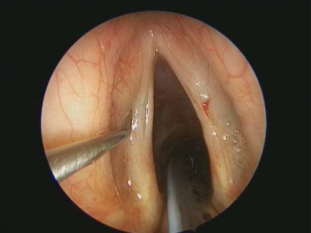
Sulcus vocal intraoperative 9 21 2011
 |
 |
 |
|
examination with microscope during laryngoscopy |
palpation of vocal fold during direct laryngoscopy |
palpation of vocal fold during direct laryngoscopy |
video
Sulcus Injection
References
Ford CN, Inagi K, Khidr A, Bless DM, Gilchrist KW; . Sulcus vocalis: a rational approach to diagnosis and management. Ann Otol Rhinol Laryngol. 1996;105:189-200
Sunter AV, Yigit O, Hug GE, Alkan Z, Kocak l, Buyuk Y; Histopathologic Characteristics of Sulcus Vocalis; Otolaryngol Head Neck Surg. 2011 Aug;145(2):264-9.
Giovanni A, Chanteret C, Lagier A; Sulcus Vocalis: A Review; Eur Arch Otorhinolaryngol. 2007 Apr;264(4):337-44.
Mallur PS, Gartner-Schmidt J, and Rosen CA: Voice Outcomes Following the Gray Minithyrotomy. Annals of Otology, Rhinology & Laryngology 121(7): 490-496
Itoh T, Kawasaki H, Morikawa I, and Hirano M: Vocal fold Furrows. A 10-year review of 240 patients. Auris Nasus larynx. 1983;10 Supple:S17-26