


click on image above to enlarge; advance with cursor over lateral border
Return to: Posterolateral Neck Dissection or Case Example Posterolateral Neck Dissection

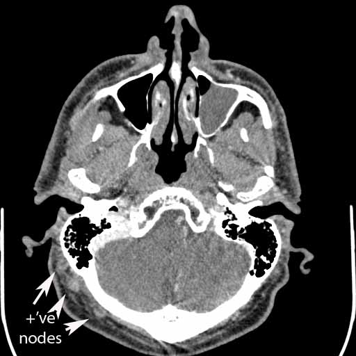
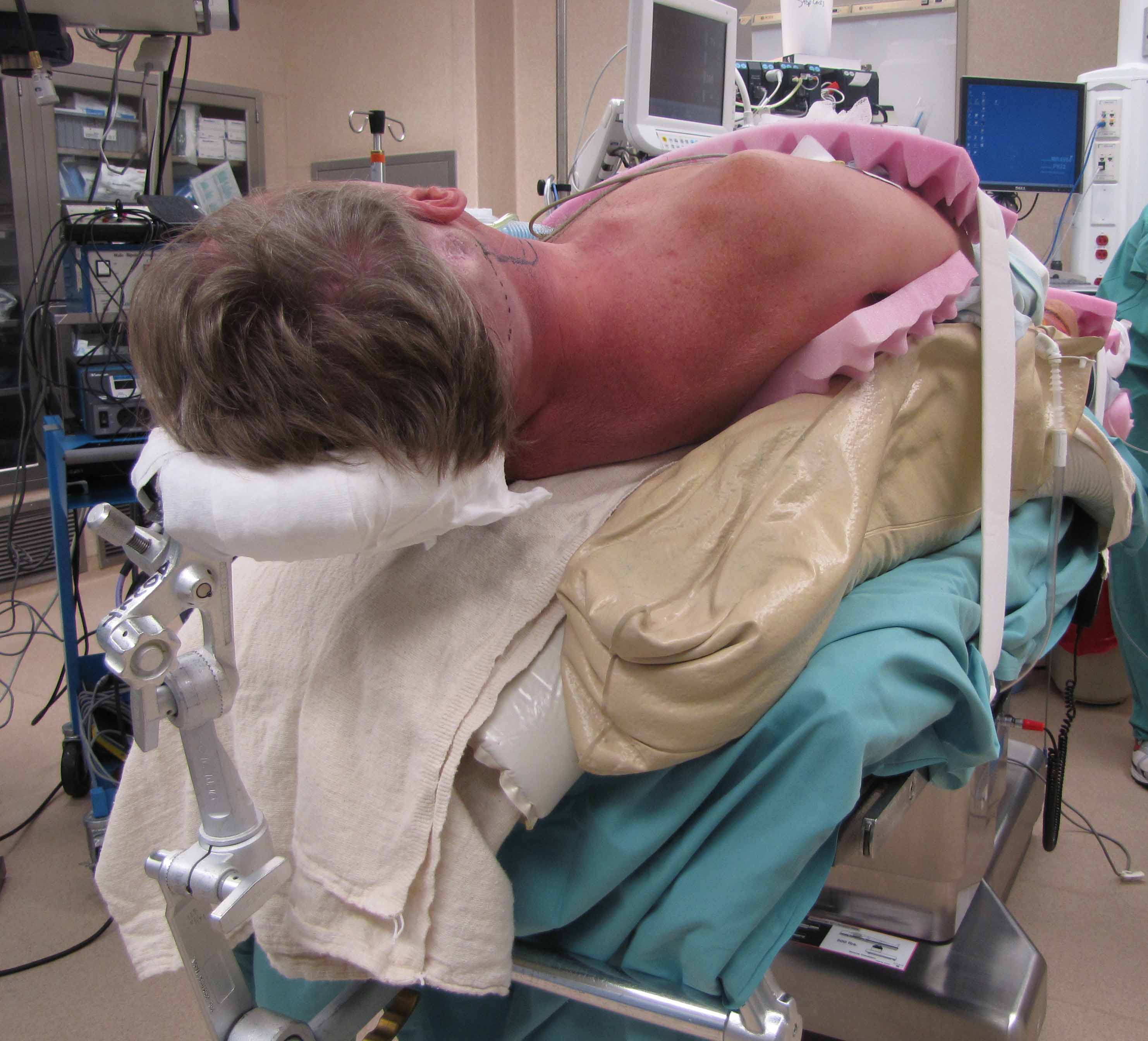
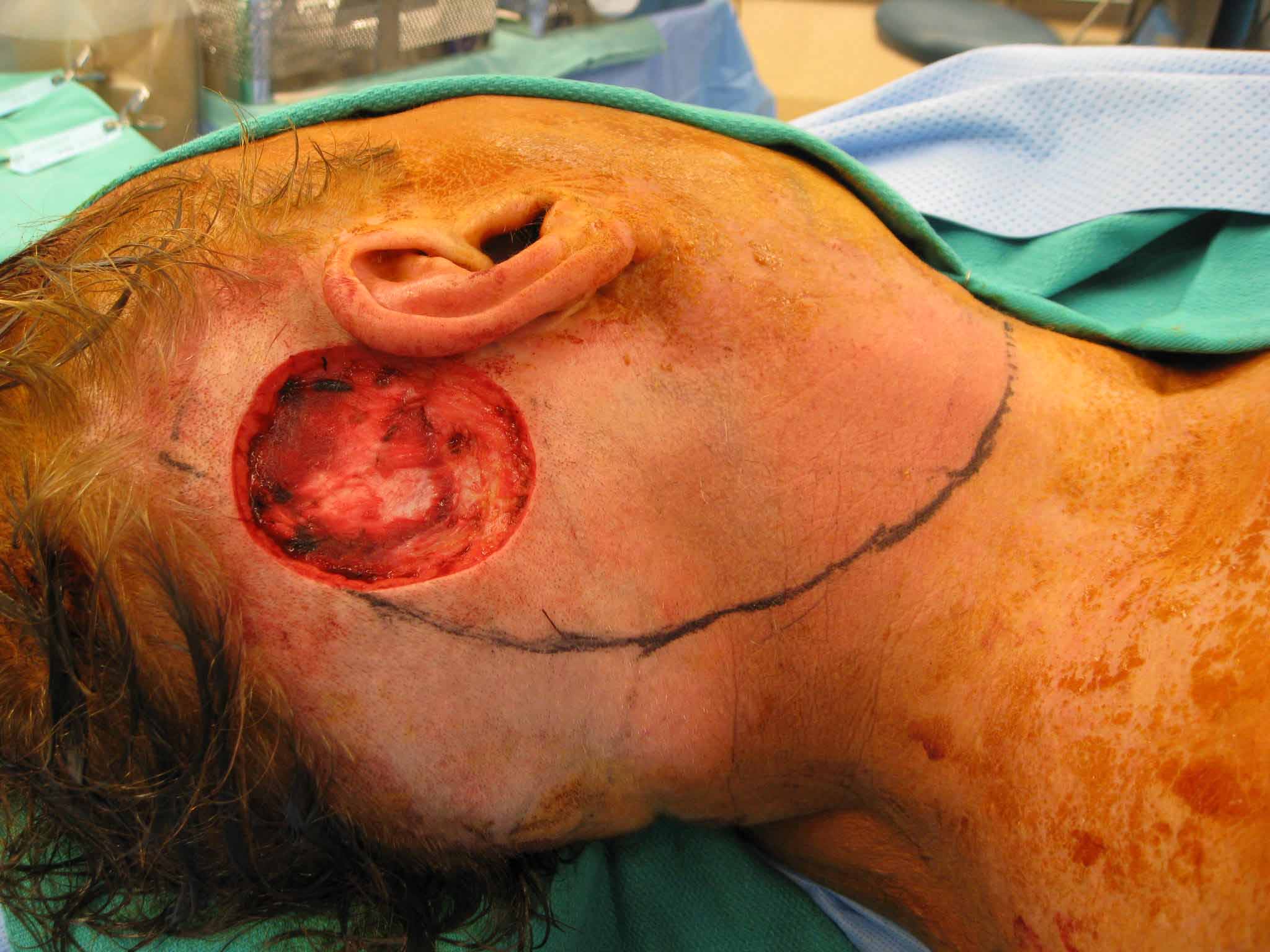
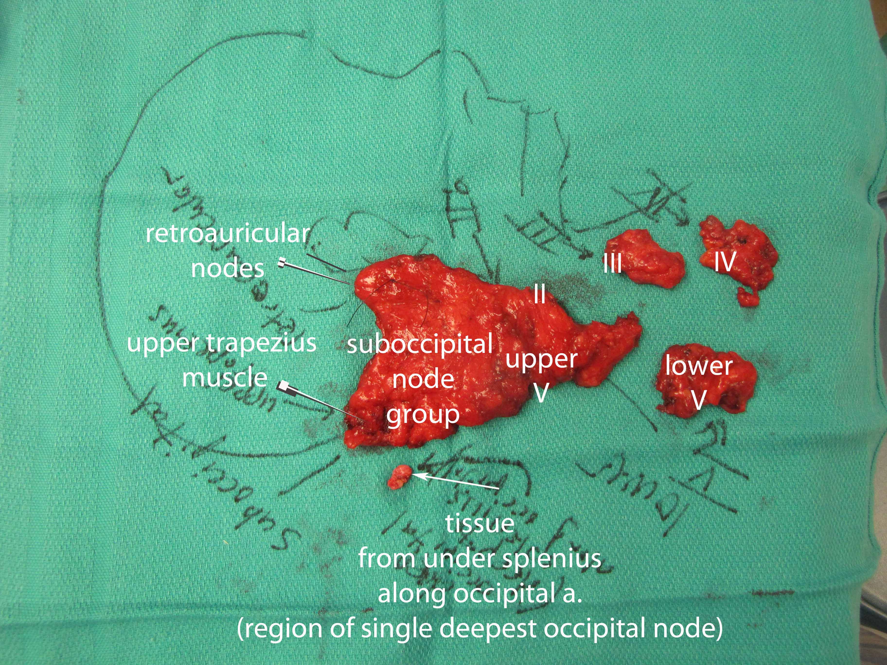
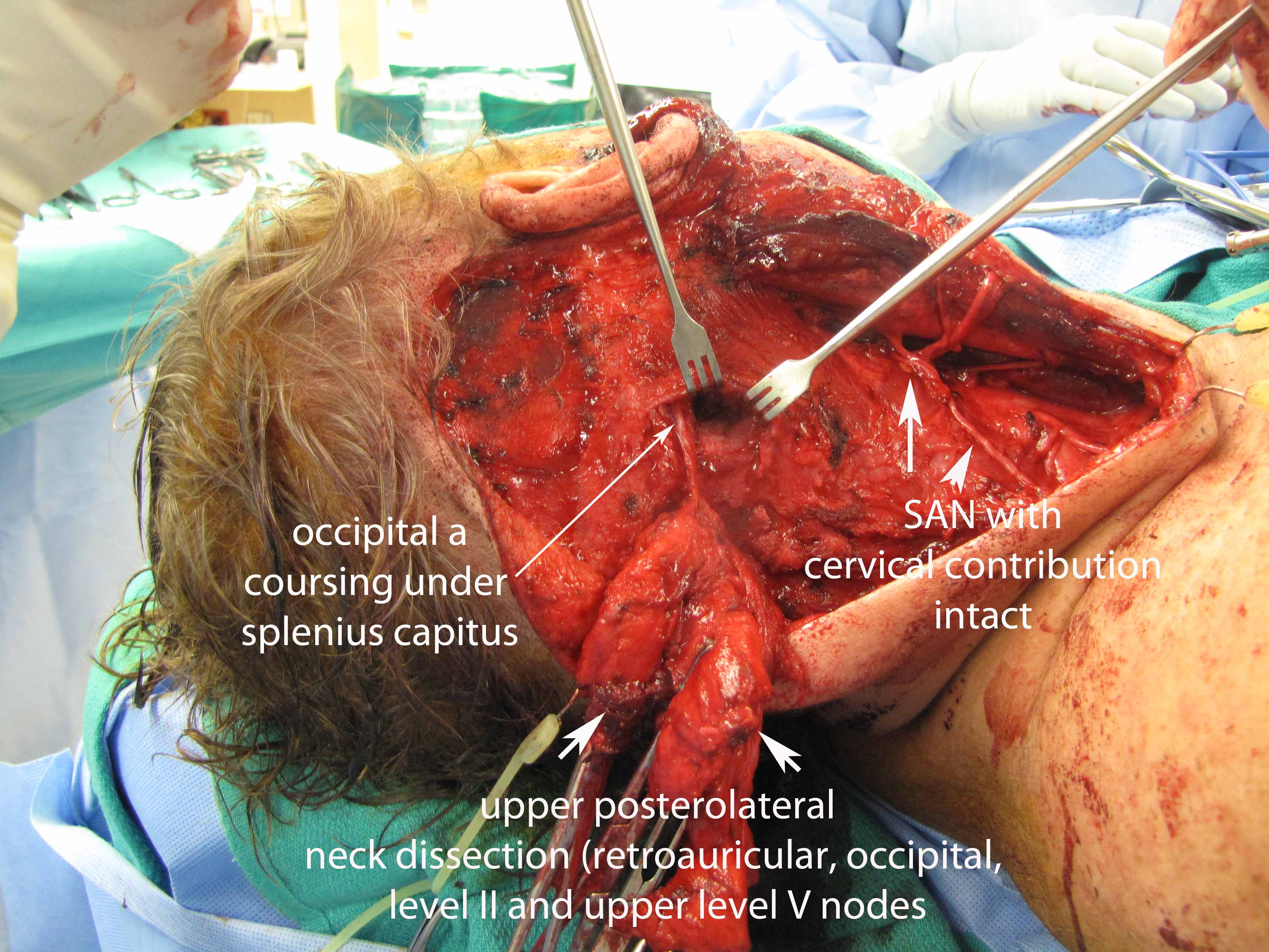
51 yo with ulcerated melanoma of scalp/post-auricular region with 20 mm depth invasion with lymphovascular spread. PET uptake in post-auricular and suboccipital region initial ascribed to inflamation from excisional biopsy. CT showed nodes confirmed ultrasound guided FNA as positive for melanoma. Posterolateral neck dissection showed 5 out of 32 nodes positive for metastases. All 5 positive nodes were located in the post-auricular region and occipital chain along the nuchal line.


Anatomy
Erb's Point (punctum nervosum)
- Location: at angle between posterolateral border of sternocleidomastoid muscle and clavicle
- Formed by C5, C6 nerve roots, cutaneous branches of cervical plexus (lesser occipital nerve, great auricular nerve, transverse cervical nerve, supraclavicular nerve)
- Electrical stimulation causes contractions of biceps, deltoid, brachioradialis, brachialis
- Clinical Significance (Erb's Palsy):
- injury commonly occurs at birth or from a fall onto the shoulder
- normally injury to C5 and C6
- symptoms include paralysis of biceps, brachialis, and coracobrachialis (through musculocutaneous nerve) and brachioradialis (through radial nerve) and deltoid (through axillary nerve)
- patient's arm hangs at side with hand rotated medially, “waiter's tip hand”
Phrenic Nerve (C3-C5)
- Contains motor, sensory, and sympathetic nerve fibers
- Provide only motor innervation to the diaphragm and sensation to the central tendon
- In the thorax, supplies mediastinal pleura and pericardium
- Both phrenics run along the anterior scalene muscle
- right phrenic – passes over brachiocephalic artery, posterior to subclavian vein, then crosses root of right lung anteriorly and leaves thorax by passing through vena cava hiatus opening in the diaphragm at the level of T8, passing over right atrium
- left phrenic – passes over pericardium of left ventricle and pierces diaphragm separately
- Clinical Significance:
- pain in regions innervated by phrenic nerve are often referred to the shoulder (ex: Kehr's sign – pain referred to right shoulder from abscess underneath right diaphragm)
Internal Jugular Vein
- Made of the inferior petrosal sinus and the sigmoid sinus
- Begins in the posterior compartment of the jugular foramen
- Drains anterior branch of retromandibular vein, facial vein, lingual vein
Occipital Artery
- Arises from external carotid artery opposite facial artery
- Passes below posterior belly of digastric to occipital region
- Supplies blood to back of scalp and sterno-mastoid muscles
|
Muscle |
Origin |
Insertion |
Innervation |
Function |
|
Sternocleidomastoid (sternal head) |
Upper part anterior surface manubrium of sternum |
Lateral one-half of superior nuchal line |
Spinal accessory nerve and branches from anterior rami of C2 to C3 |
Tilt head towards shoulder on ipsilateral side rotating to turn face to opposite side, if act bilaterally draw head forward |
|
Sternocleidomastoid (clavicular head) |
Superior surface of medial one-third of clavicle |
Lateral surface of mastoid process |
|
|
|
Trapezius |
Super nuchal line; external occipital protuberance; ligamentum nuchae; spinous processes of vertebrae CVII to TXII |
Lateral one-third of clavicle; acromion; spine of scapula |
Motor – spinal accessory nerve; proprioception – C3 and C4 |
Assists in rotating scapula during abduction of humerus above horizontal; upper fibers – elevate, middle fibers – adduct, lower fibers – depress scapula |
|
Splenius capitis |
Lower half of ligamentum nuchae; spinous processes of vertebrae CVII to TIV |
Mastoid process; skull below lateral one-third of superior nuchal line |
Posterior rami middle cervical nerves |
When act together draw head backwards; individually draw and rotate head to ipsilateral side |
|
Levator scapulae |
Transverse processes of C1 to C4 |
Upper part of medial border of scapula |
C3, C4, and dorsal scapular nerve (C4, C5) |
Elevates scapula |
|
Posterior scalene |
Posterior tubercles of transverse processes of vertebrae CIV to CVI |
Upper surface of rib II |
Anterior rami of C5 to C7 |
Elevation of rib II |
|
Middle scalene |
Transverse processes of vertebrae CII to CVII |
Upper surface of rib I posterior to the groove for the subclavian artery |
Anterior rami of C3 to C7 |
Elevation of rib I |
|
Anterior scalene |
Anterior tubercles of the transverse processes of vertebrae CIII to CVI |
Scalene tubercle and upper surface of rib I |
Anterior rami of C4 to C7 |
Elevation of rib I |
|
Omohyoid |
Superior border of scapular medial to scapular notch |
Inferior border of body of hyoid bone |
Ansa cervicalis; anterior rami of C1 to C3 |
Depress the hyoid bone |
Lymph Node Levels
- Level I : below myohyoid to hyoid bone anteriorly
- Level Ia : submental
- Level Ib : submandibular
- Level II : jugulodigastric (base of skull to hyoid)
- Level III : deep cervical (hyoid to cricoid)
- Level IV : Virchow (cricoid to clavicle)
- Level Va : accessory spinal (posterior triangle) : superior half
- Level Vb : accessory spinal (posterior triangle) : inferior half
- Level VI : prelaryngeal / pretracheal / Delphian node
- Level VII : superior mediastinal (between CCAs, below top of manubrium)
References
Drake R; Vogl W; Mitchell A. (2005). Gray's anatomy for students. Posterior triangle of the neck (pp 919 - 927). Canada: Elsevier.