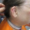
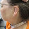
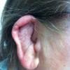
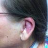
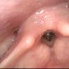
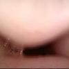
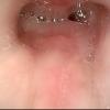
click on image above to enlarge; advance with cursor over border
return to: Relapsing Polychondritis for Otolaryngologists (ENT)
Case History
July 28 2009 61 yo female with progressive stridor beginning three years previously was first evaluated at the UIHC on July 28, 2009 with subglottic narrowing identified as a likely due to small stature (5'4"), LPR (reflux to larynx and airway), compounded by diabetes and a history of weight loss from 210 pounds (1 1/2 years ago) to 175 pounds. She had an uncomplicated remote (12 years ago) intubation for a brief surgical procedure. She identified continued reflux symptoms (gas, 'burping up food', frequent throat clearing) continued despite a careful diet and use of once-a-day Prevacid.
Episodes of shortness of breath had responded to steroids - requiring them '50% of the time over the past 7 months". A pulmonary medicine evaluation at another institution revealed what were felt to be normal lungs. CT of neck 2-2009 showed narrowing in the subglottis.
Flexible fiberoptic transnasal laryngoscopy performed after instillation of lidocaine with phenylephrine into the nostril showing some mild reflux changes to the supraglottic larynx with some blunting and a bit of Reinke's space edema and subglottic narrowing approximately 1.5 cm below the vocal folds. Evaluation included a negative rheumatoid factor, ANA, C-ANCA and P-ANCA, and urinalysis.
She was admitted to the pulmonary ICU on high dose steroids with Jackson metal laryngeal dilators at her bedside (and to accompany her throughout the hospital).
July 29 2009 Operative intervention the next day with microdirect laryngoscopy with tracheoscopy, kenalog 10 (1 part kenalog 40, 3 parts 1% with 1:100,000) injection to 0.8 cc to subglottis, radial cuts at 3:00, 9:00 and 12:00 with biopsy (stromal fibrosis focal chronic inflammation) at 9:00, dilation (under jet anesthesia) to 36 Jackson laryngeal dilator, then intubation with 4-0 MLT with use of CRE Pulmonary (controlled radial expansion dilation) to 1.5 atmospheres (approx 15mm) with 4-0 MLT tube in place (diameter of expansion greater than 15 mm); Cervial and flexible fiberoptic esophagoscopy was unremarkable
August 1 2009 Discharged home breathing well (the best in years).
August 7 2009 Steroid taper completed - with rapid return of shortness of breath warranting
August 9 2009 Readmission to SICU through the emergency room with stridor
August 13 2009 Operative intervention with microdirect laryngoscopy (photos only) and tracheotomy
September 2010 Referral to rheumatology for onset of ear pain and swelling as well as chest discomfort - felt to reflect costochrondritis with clinical diagnosis of relapsing polychondritis warranting treatment (with response) with steroids and CellCept. (prednisone 20 mg daily, alendronate 70 mg weekly (bisphosphonate), and mycophenolate mofetil 500 mg PO BID for 14 days, then 1000 mg PO BID (CellCept).
October 21, 2010 Evaluation in Otolaryngology (see photos above)
Comment: The favorable and reproducible response to systemic steroids were the only atypical feature at the time of presentation that may have directed the initial diagnosis to the uncommonly considered Relapsing Polychondritis. Endoscopic findings at the time of the first microdirect laryngoscopy were also atypical for post-traumatic subglottic narrowing (no defined scar band), reflux disease (minimal inflammation) and Wegener's granulomatosis (no active granulation tissue).
Relapsing polychondritis
- Rare inflammatory disease resulting in episodic inflammation of cartilaginous structures throughout the body
- The etiology of the this disease is unknown
- M = F
- Peak onset is 4th to 5th decade
- Only 14% of pts present with airway sx but up to 55% will eventually develop them
- Most commonly affected: ears, nose, eye, larynx, bronchi, costal cartilage, and articular joints.
- 25-35% have a preceding autoimmune disorder
The diagnostic criteria for RP were initially established by McAdam (1976) to include three of the following 6 features:
- bilateral auricular chondritis
- nasal chondritis
- respiratory tract chondritis
- ocular damage
- nonerosive seronegative inflammatory polyarthritis
- audiovestibular damage
Damiani and Levine (1979) revised McAdam’s criteria to permit the diagnosis with:
- three McAdam criteria (as above) or
- one McAdam criterion and positive histology or
- two McAdam criteria and response to corticosteroids or dapsone.
Management
- In additional to medical management directed by Rheumatology, coordination of consultations with other services (such as Pulmonary medicine) is usually assigned to a single care giver (most often Rheumatology) - to ensure cohesive and comprehensive care.
- Otolaryngology interventions center around upper airway management supplemented by Otologic issues. Assessment with an audiogram and provision of hollow ear molds to maintain patency to the collapsed cartilaginous ear canal may be useful.
References
Damiani JM, Levine HL. Relapsing polychondritis: report of ten cases. Laryngoscope 1979; 89: 929--946.Int J Dermatol. 2009 Apr;48(4):356-62.
McAdam LP, O’Hanlan MA, Bluestone R,et al. Relapsing polychondritis: prospective study of 23 patients and a review of the literature. Medicine (Baltimore)1976;55:193--215.
Watkins S, Magill JM Jr, Ramos-Caro FA.Int J Dermatol. Annular eruption preceding relapsing polychondritis: case report and review of the literature. 2009 Apr;48(4):356-62.
Flint, PW et al. Cummings Otolaryngology: Head and Neck Surgery, 5th ed. Chap 64. Mosby Elsevier. Philadelphia, PA. 2010
Sato T, Yamano Y, Tomaru U et al: Serum level of soluble triggering receptor expressed on myeloid cells--1 as a biomarker of disease activity in relapsing polychondritis. Modern Rheumatology Feb 13 2013
