


click on image above to enlarge; advance with cursor over border
see also: Case Example Full Thickness Skin Graft (subsequent reconstruction with full thickness skin graft with long term f/u)
return to: Melanoma (Evaluation and Management)
Case Example
General anesthesia was induced and he was intubated with the tube secured in left oral commissure. The table was turned 180 degrees. His head of bed was elevated. 2 cc of plain 1:100,000 epinephrine (no lidocaine) was injected into his right temple region. Following this, the patient was prepped and draped in a sterile fashion with ground pad for Parsons McCabe facial nerve stimulator placed.
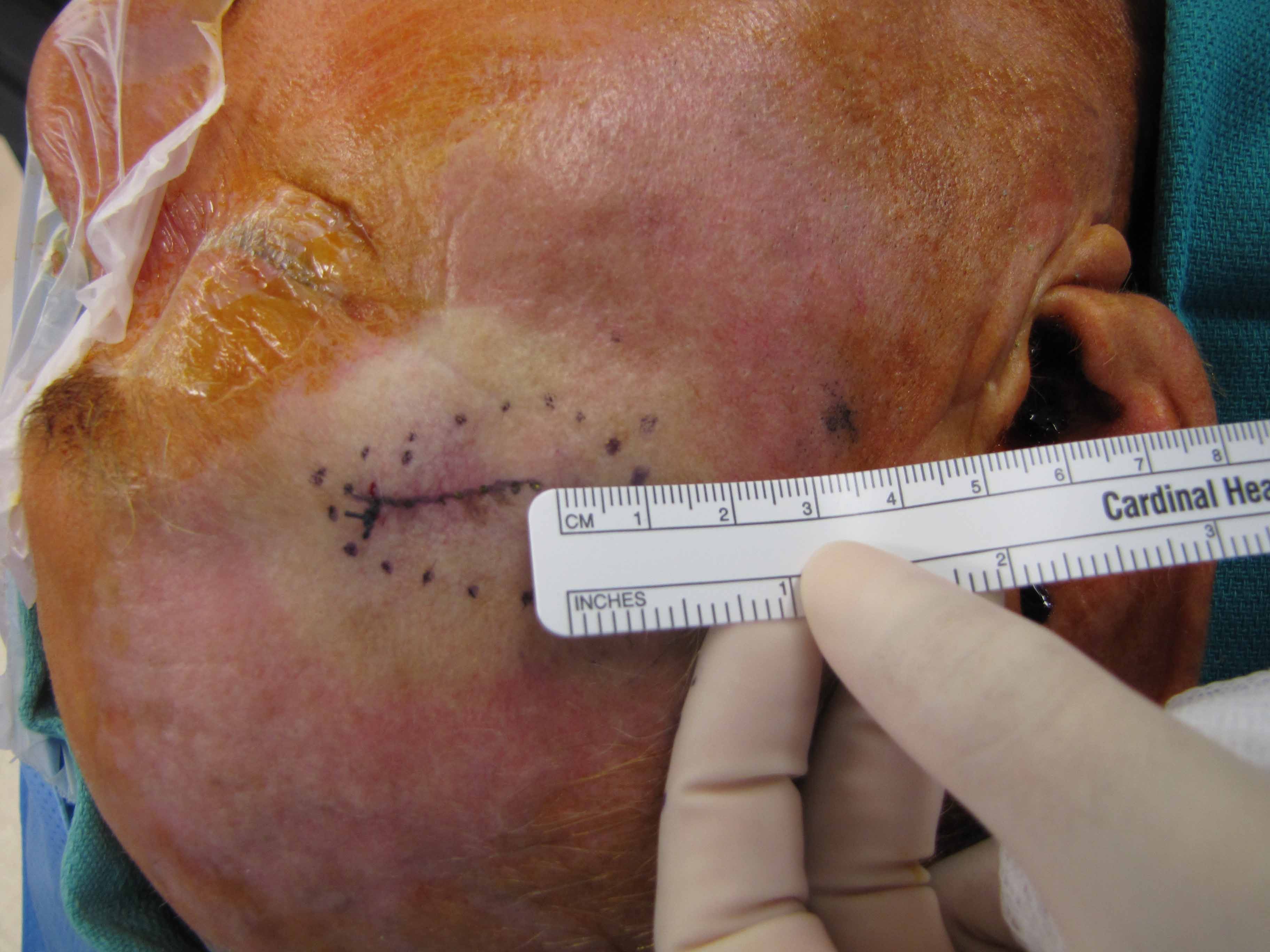
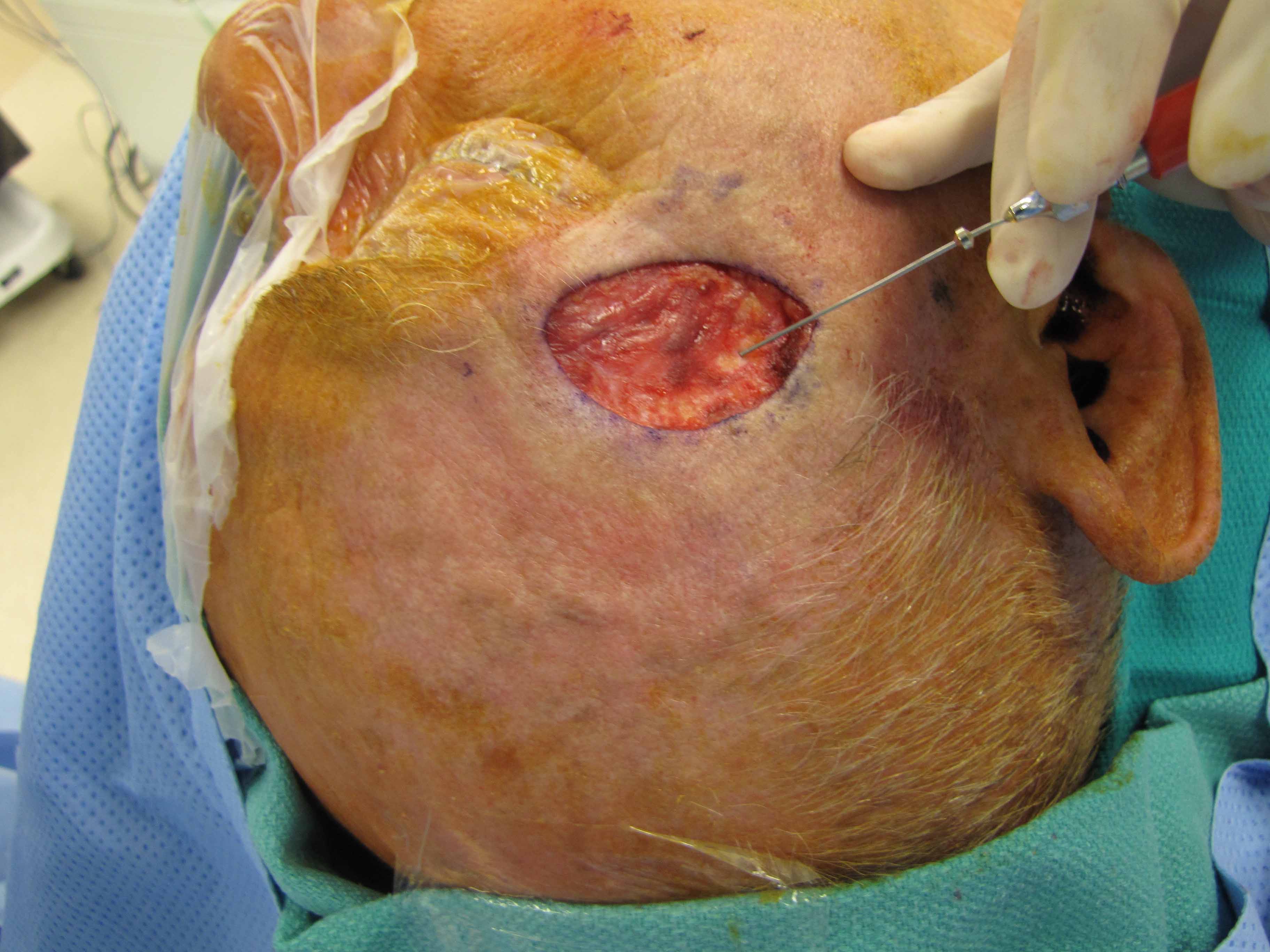
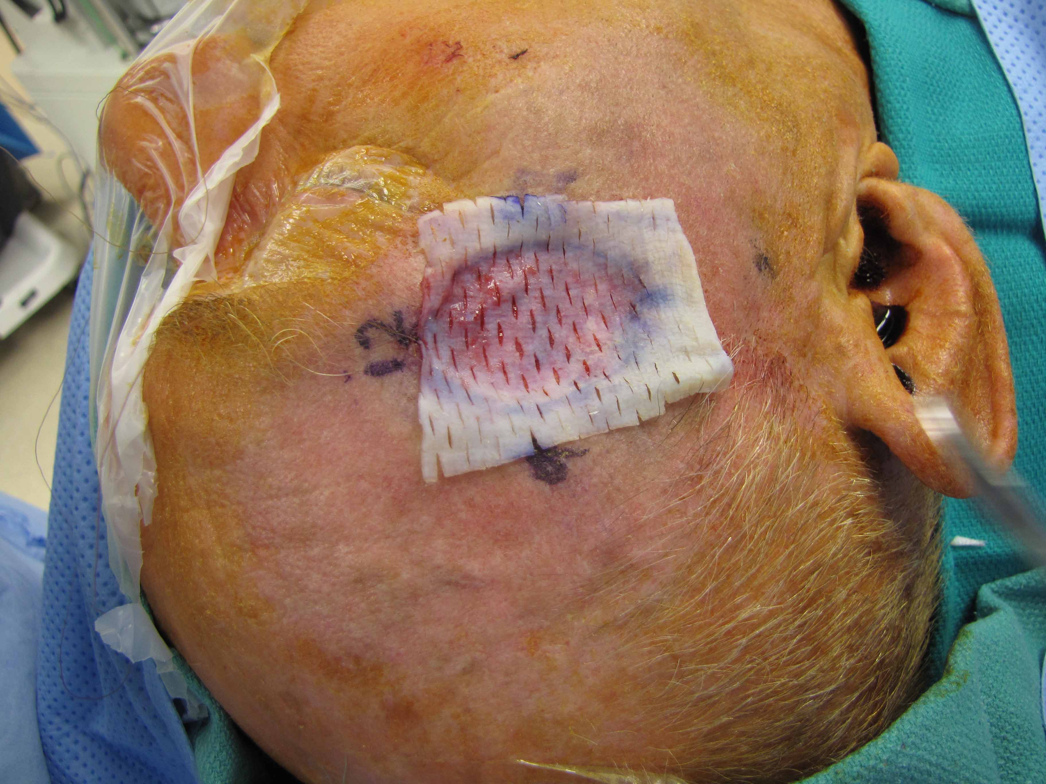
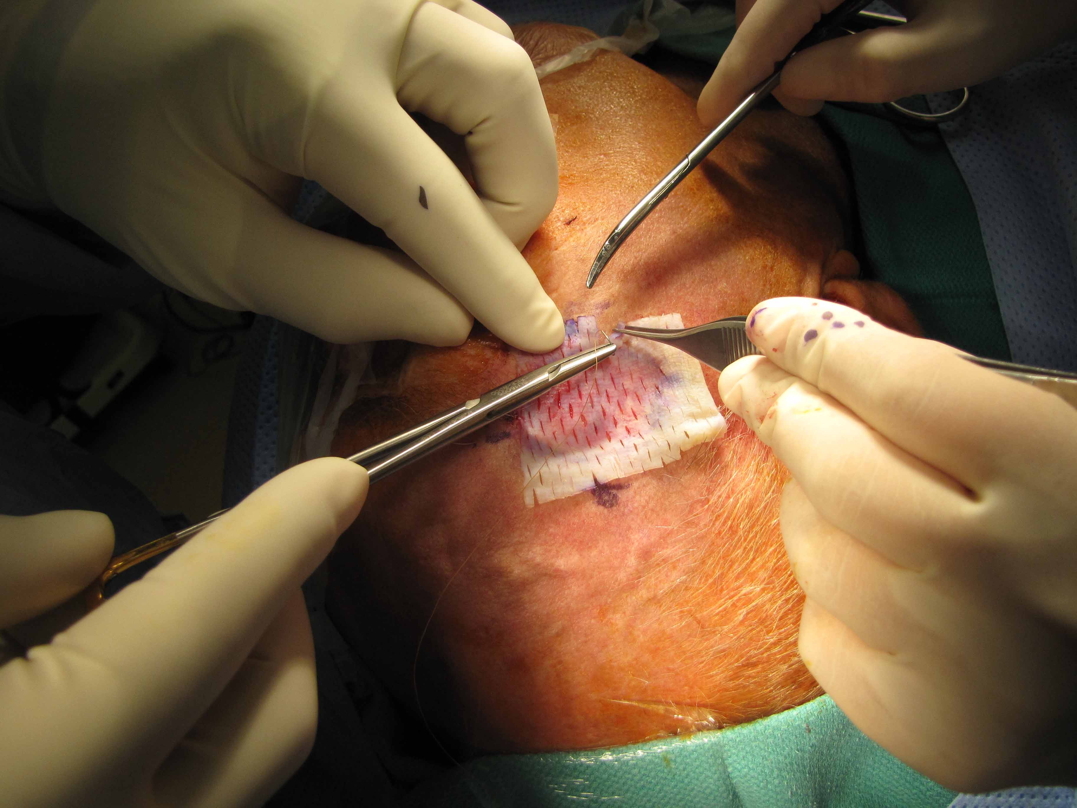
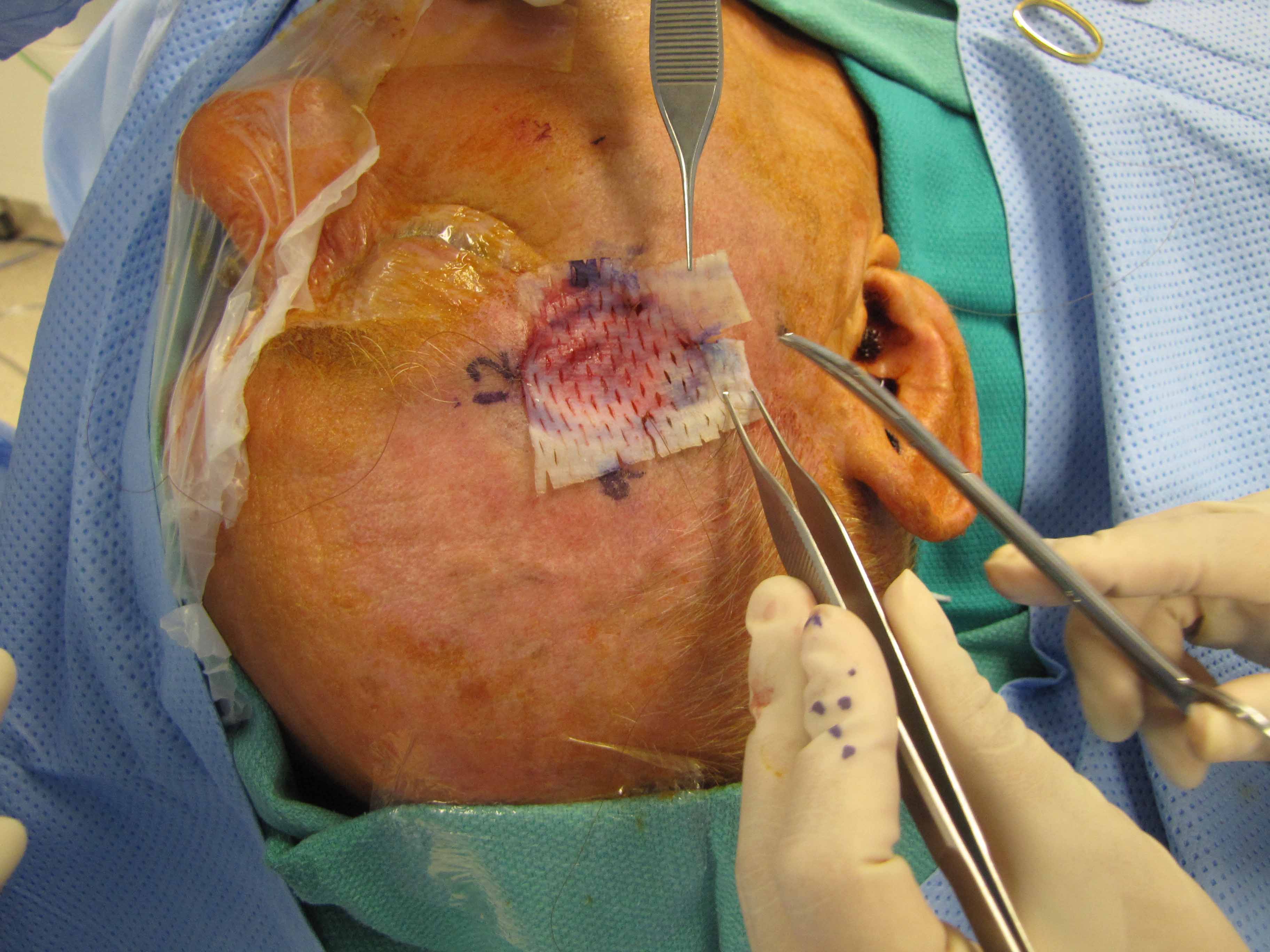
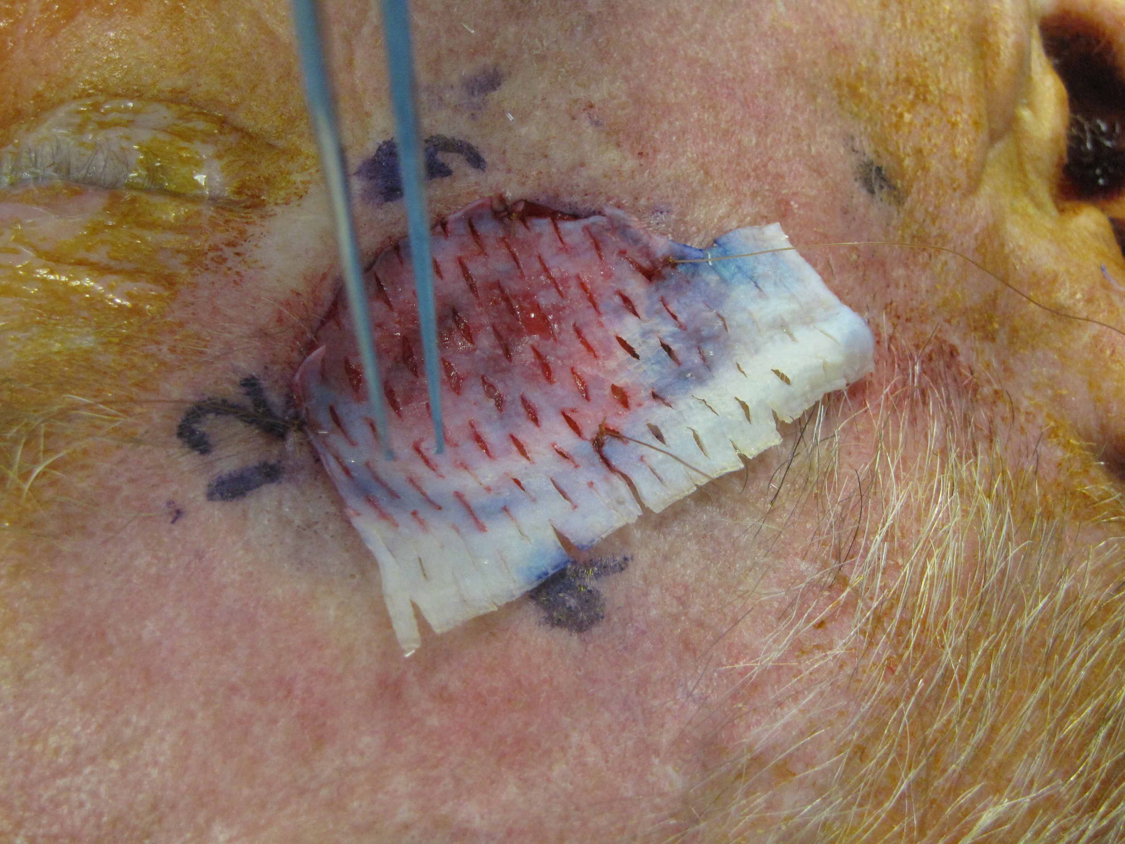
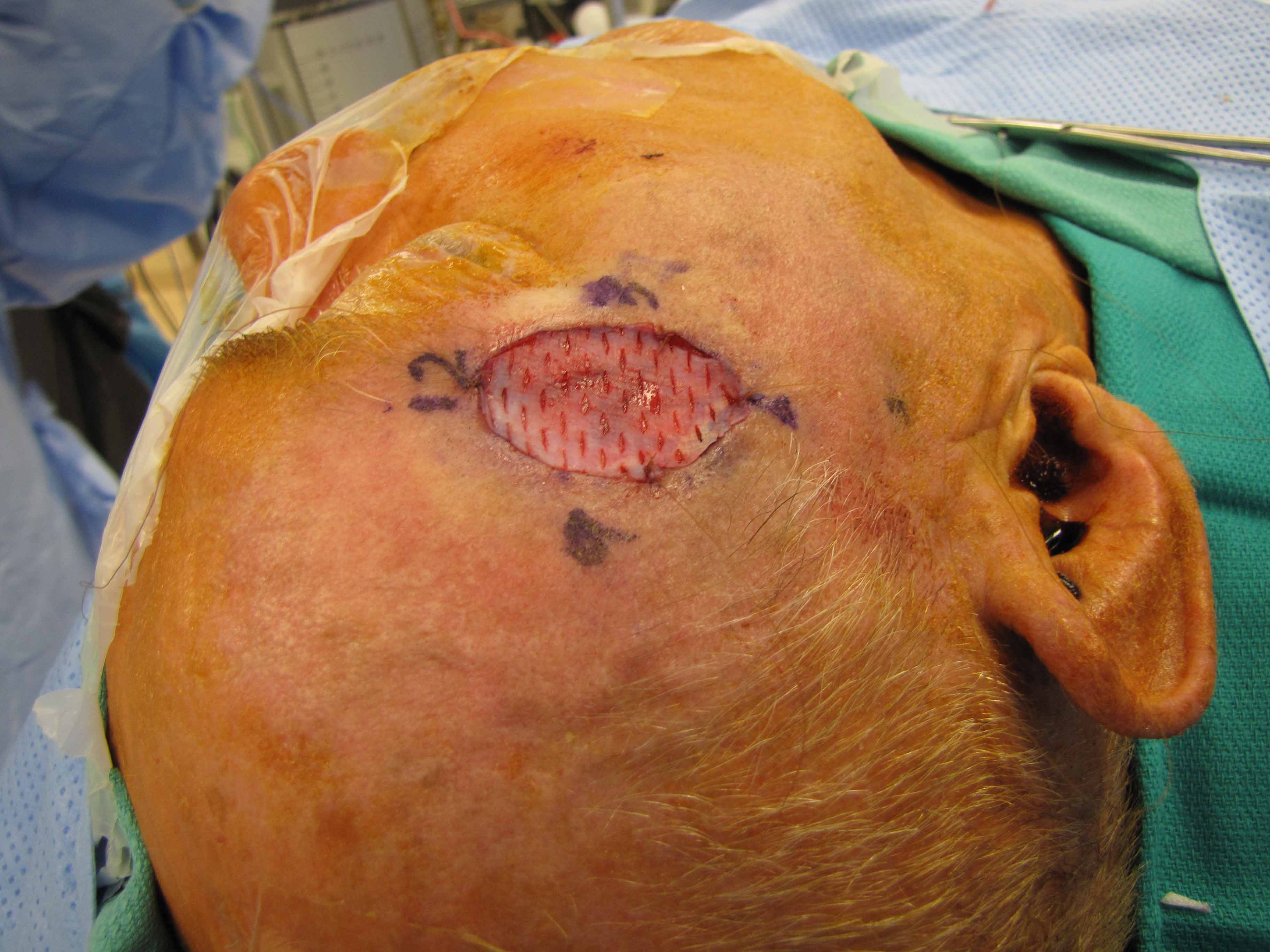
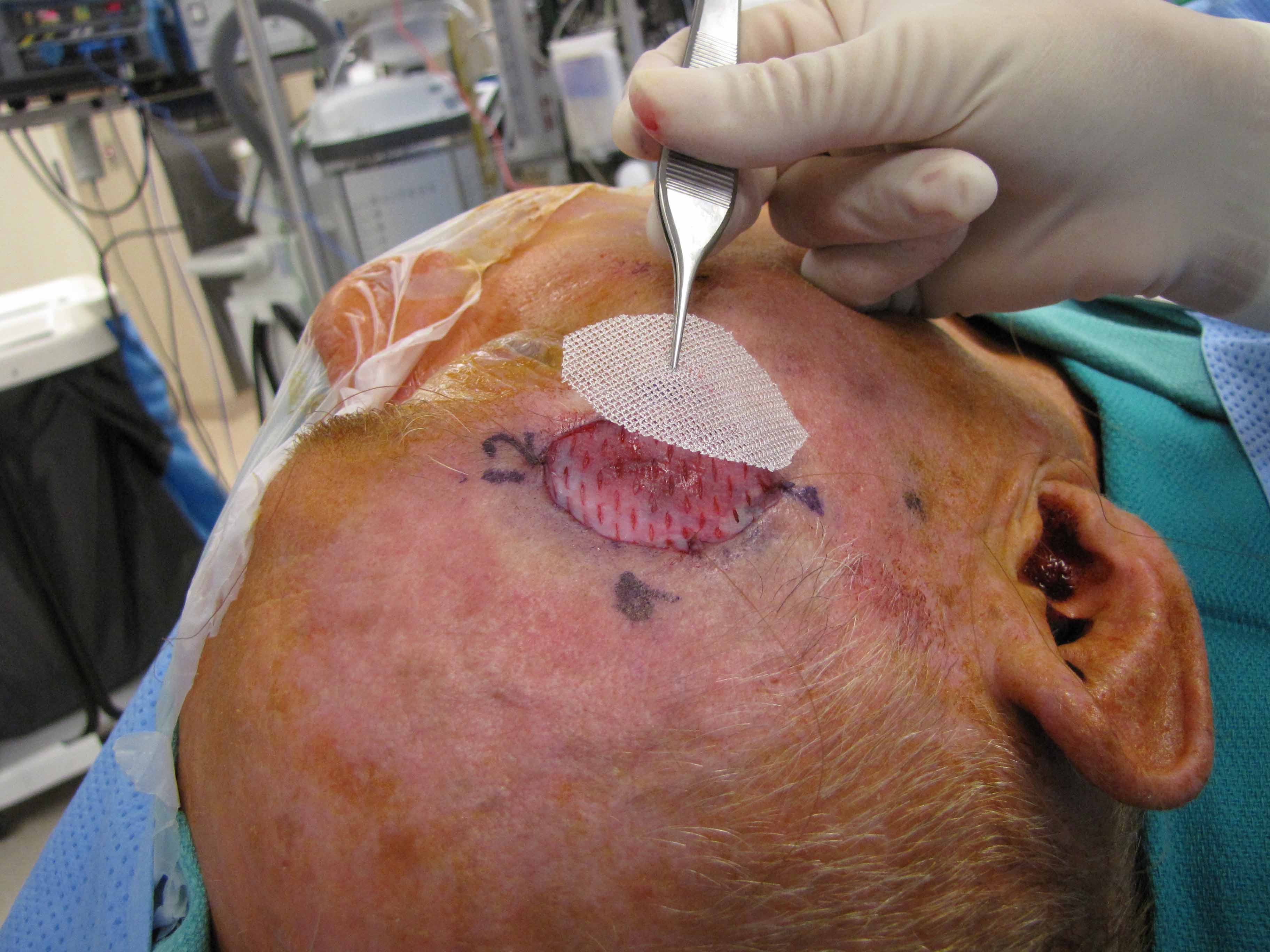
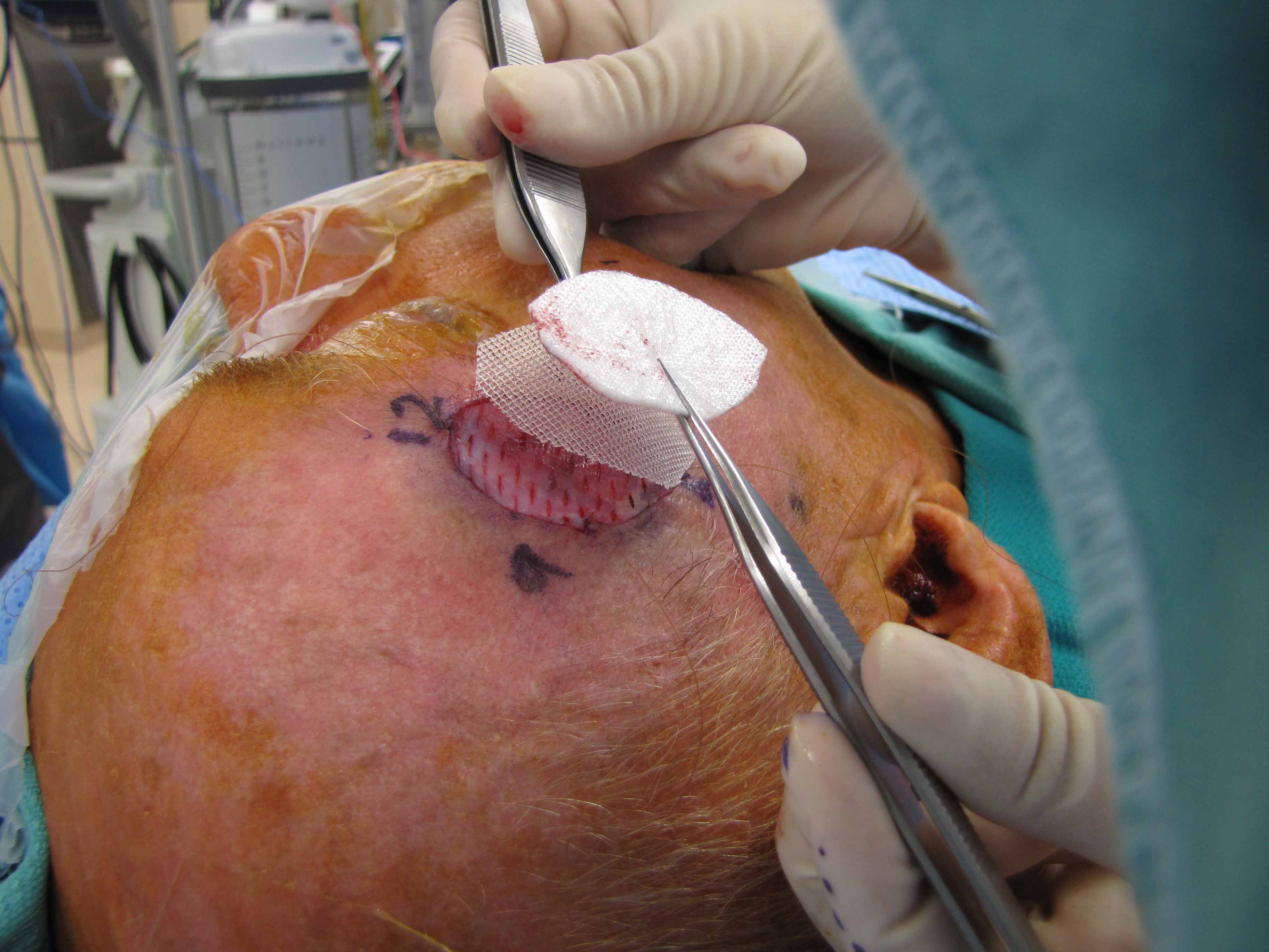
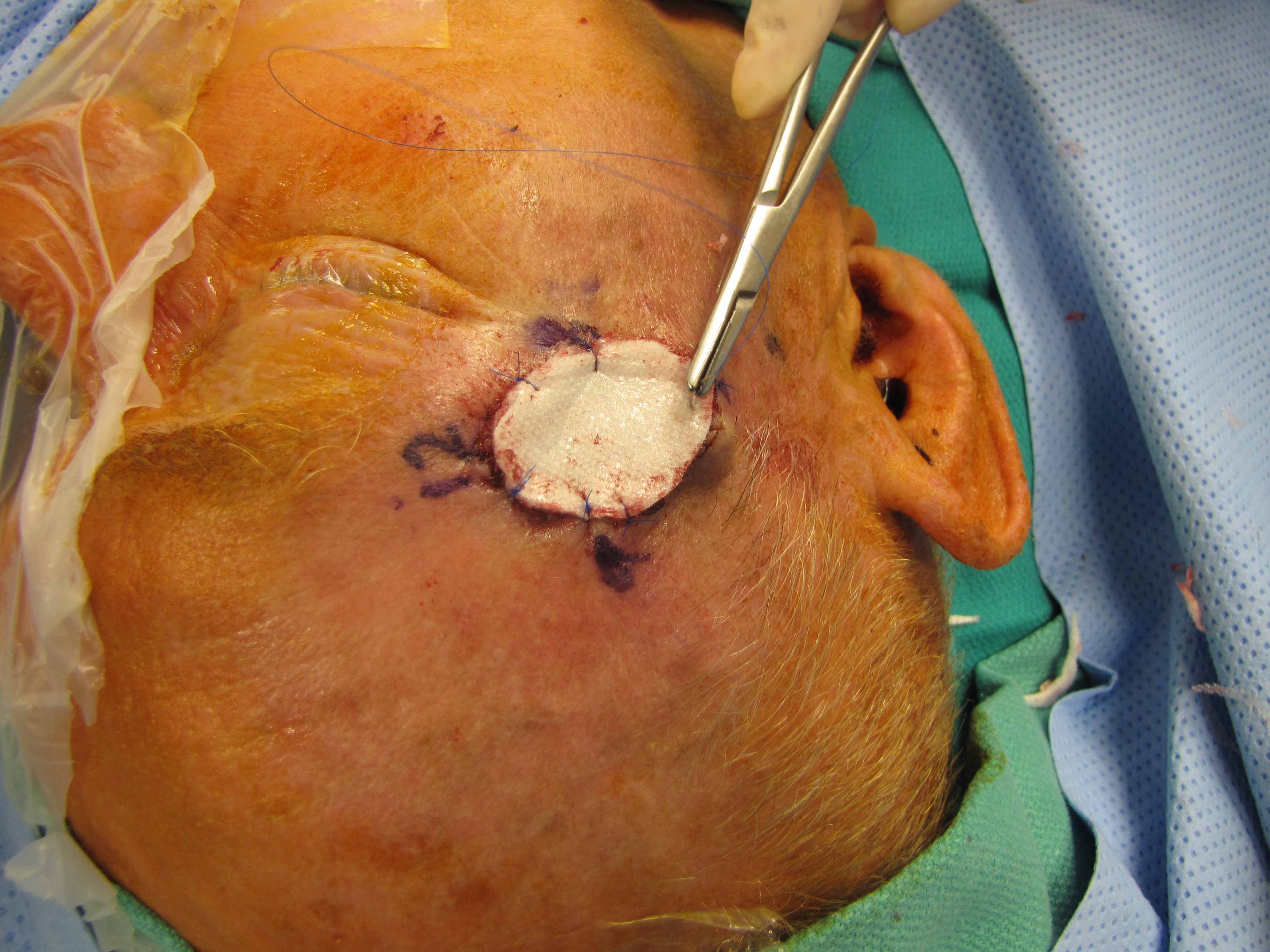
An elliptical incision was marked surrounding the previous scar to widely encompass with 1 cm + margins about the central portion of the scar - yielding a margin was 1 cm laterally and 0.5 cm medially close to his eye. After this margin was marked, the 15 blade was used to make an incision through the dermis. Careful dissection from a lateral to medial approach permitted identification of the frontal branch of the facial nerve employing the Parsons-McCabe facial nerves stimulator. Dissection was performed in a plane lateral to the nerve and the fascia of the orbicularis oculi. The excision was measured at 3.5 x 2.5 cm. Hemostasis was achieved with pressure. There was no cautery that was used. Following this, the porcine graft was measured and was sutured into the area with 5-0 chromic. The bolster was then applied after placing bacitracin opthalmic ointment over the porcine graft, then one layer of Adaptic, then 2 layers of Telfa. The dressing was then sutured with 5-0 Prolene circumferentially in simple interrupted fashion.