-
A malignant tumor of the cartilage
-
Most often seen as a mass of soft tissue with varying degrees of calcification, indistiguishable border with the bone of origin
-
Often greater than 4 cm at presentation (Limaiem et al. 2023).
-
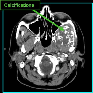
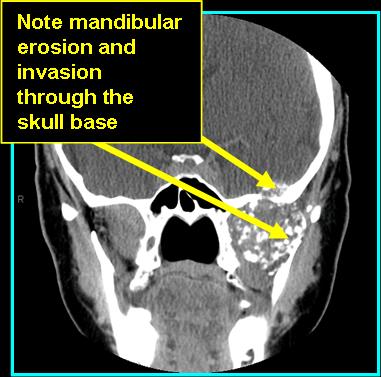
On CT:
-
Non-contrast erosion of surrounding bone, ringed or crescent calcification in low grade lesions, amorphous calcification in high grade
-
Contrast shows heterogenous enhancement, ill-defined boders
-
-
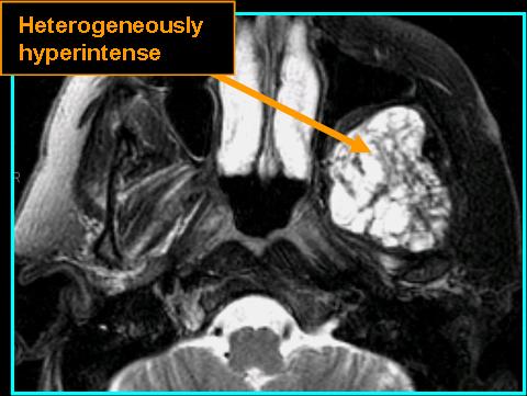
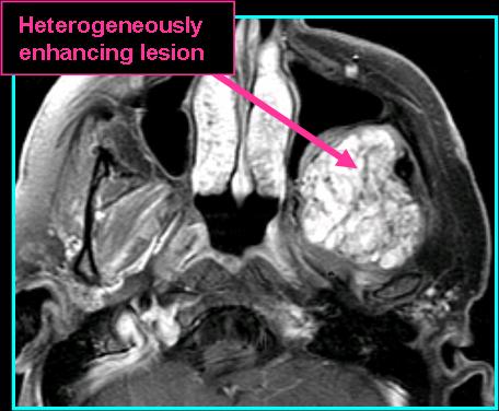
On MR:
-
T1 shows homogenous isointense signal, bone invasion is well-noted as well as marrow replacement
-
T2 shows high signal with possible surrounding edema
-
T1 post-contrast shows varying enhancement, more with higher grade lesion
-
-
Most cases are sporadic, most commonly seen above the age of 50 years (Limaiem et al. 2023).
-
The second most common bony malignancy (Weinschenk et al. 2021 and Chow 2018).
-
Late-recurrences not uncommon; histological grade is the most important predictor (Limaiem et al. 2023).
![]()
![]()
![]()
References
Limaiem F, Davis DD, Sticco KL. Chondrosarcoma. [Updated 2023 Aug 14]. In: StatPearls [Internet]. Treasure Island (FL): StatPearls Publishing; 2023 Jan-. Available from: https://www.ncbi.nlm.nih.gov/books/NBK38132/
Weinschenk RC, Wang WL, Lewis VO. Chondrosarcoma. J Am Acad Orthop Surg. 2021;29(13):553-562. doi:10.5435/JAAOS-D-20-01188
Chow WA. Chondrosarcoma: biology, genetics, and epigenetics. F1000Res. 2018;7:F1000 Faculty Rev-1826. Published 2018 Nov 20. doi:10.12688/f1000research.15953.1