-
Also known as developmental oral cavity cyst or ectodermal inclusion cyst
-
Congenital cyst resulting from inclusion of surface ectoderm (Shareef et al. 2022).
-
Dermoid cyst contains epithelial elements plus dermoid substructure including dermal appendages
-
Epidermoid cyst is derived from ectoderm only (Quintanilla-Dieck et al. 2018).
-
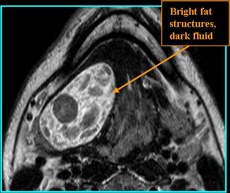
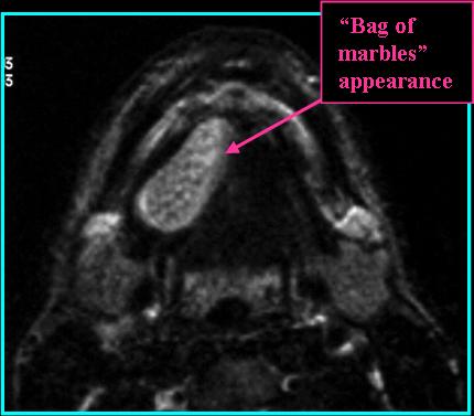
Dermoid appears as a cystic, well-defined mass with fatty, fluid, or mixed components; epidermoid is only fluid-filled
-
7% of dermoid cysts present in the head and neck region (Quintanilla-Dieck et al. 2018).
-
The lateral eyebrow area is the most common site in the head and neck region, followed by the floor of the mouth (Santos et al. 2020)
-
-
Present from birth, slow-growing, male to female ratio is 2.5:1 for dermoid cysts and 1.6:1 for epidermoid cysts (Santos et al. 2020).
-
On CT: Low density, a variety of internal appearances depending on composition, wall may enhance with contrast
-
On MR: On T1 fatty elements appear bright, fluid dark; on T2 fat is dark and fluid is bright; on T1 post-contrast thin wall enhancement possible
![]()
References
Shareef S, Ettefagh L. Dermoid Cyst. In: StatPearls. Treasure Island (FL): StatPearls Publishing; August 29, 2022.
Quintanilla-Dieck L, Penn EB Jr. Congenital Neck Masses. Clin Perinatol. 2018;45(4):769-785. doi:10.1016/j.clp.2018.07.012
Santos HB, Rolim LS, Barros CC, Cavalcante IL, Freitas RD, Souza LB. Dermoid and epidermoid cysts of the oral cavity: a 48-year retrospective study with focus on clinical and morphological features and review of main topics. Med Oral Patol Oral Cir Bucal. 2020;25(3):e364-e369. Published 2020 May 1. doi:10.4317/medoral.23388