Logo for University of Iowa Health Care This logo represents the University of Iowa Health Care
Meet the Iowa Otolaryngology Research Scholar FellowClick Here

Invasive squamous cell carcinoma causing laryngeal leukoplakia

last modified on: Wed, 09/06/2017 - 11:08

(Click on a photo and use the right and left arrow keys to toggle back and forth)

Invasive squamous cell carcinoma

Invasive squamous cell carcinoma causing laryngeal leukoplakia 

Return to: Laryngeal leukoplakia white plaques on vocal cords

Go to: Verrucous squamous carcinoma causing laryngeal leukoplakia 

Invasive squamous cell carcinoma

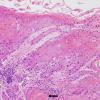
Mucosal biopsy showing a keratotic surface with infiltrating tongues and nests of malignant cells (white arrows) extending into the submucosa. Deep keratinization (black arrow) is always abnormal and should prompt one to exclude carcinoma.
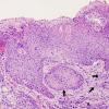
Intermediate magnification of a left true vocal cord lesion showing invading nests (black arrows) of malignant cells within the superficial submucosa.
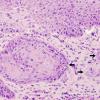
High magnification of prior biopsy shows nests of markedly atypical squamous cells (black arrows) invading into the submucosa. Note the squamous pearls, deep keratinization, and reactive appearance to the stroma.
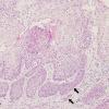
Intermediate magnfication of a frozen section (intraoperative consultation) of a laryngeal biopsy showing an expansile tumor with infiltrating buds of atypical squamous cells (black arrows) invading the submucosa.
High magnification of the prior biopsy showing irregular nests and tongues of malignant cells (black arrows) invading the superficial submucosa.
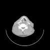
CT imaging demonstrates a large right glottic mass filling paraglottic space
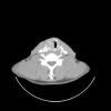
CT imaging identifies subglottic extension
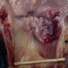
Total laryngectomy specimen showing squamous cell carcinoma involving right vocal cord, anterior commmissure and subglottis

Invasive squamous carcinoma is characterized by infiltrating tongues and nests of atypical squamous cells invading into the submucosa that are almost always accompanied by an desmoplastic stromal response. It is usually accompanied by keratinization (squamous pearl formation), increased mitotic activity, and significant cytologic atypia. Carcinoma is graded as well-differentiated, moderately differentiated, or poorly differentiated based on the degree of squamous differentiation.