updated 2024 by Piper Wenzel BS
- Rare, represent 0.7 to 4% of all posterior fossa meningiomas (Thomas et al. 2015)
- Present in 4th or 5th decade of life; more common in females (Thomas et al. 2015)
- Common presenting symptoms include hearing loss, tinnitus, and middle ear mass (Thomas et al. 2015)
- Look for permeative-sclerotic bony erosions on CT and tumor spread along dural surfaces on post-contrast T1 MR
- Typically large at presentation with poorly-defined margins and centrifugal spread, especially along dura
- Treatment involves observation with serial imaging, surgery, or radiation (Tolisano et al. 2018)
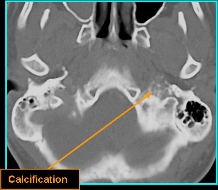
- On CT: hyperdense mass with permeative-sclerotic bony changes without contrast; high density with contrast
- CT spread patterns:
- Superolateral to middle-ear mastoid space
- Lateral to CN VII mastoid segment
- Anterior to horizontal petrous ICA canal
- Anteromedial to petrous apex
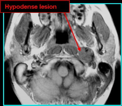
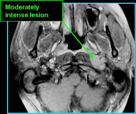
- On MR:
- Hypo- to isointense on T1 with ABSENT flow voids and rarely bright calcifications
- Hypointense on T2
- FLAIR hyperintense
- T1 post-contrast is hyperintense with good definition of spread
![]()
![]()
![]()

![]()
References
Tolisano AM, Lin K, Isaacson B. Jugular Foramen Meningioma. Otol Neurotol. 2018;39(3):e222-e223. doi:10.1097/MAO.0000000000001709
Thomas AJ, Wiggins RH 3rd, Gurgel RK. Nonparaganglioma jugular foramen tumors. Otolaryngol Clin North Am. 2015 Apr;48(2):343-59. doi: 10.1016/j.otc.2014.12.008. Epub 2015 Feb 4. PMID: 25659512.
Ota Y, Liao E, Capizzano AA, Yokota H, Baba A, Kurokawa R, Kurokawa M, Moritani T, Yoshii K, Srinivasan A. MR diffusion and dynamic-contrast enhanced imaging to distinguish meningioma, paraganglioma, and schwannoma in the cerebellopontine angle and jugular foramen. J Neuroimaging. 2022 May;32(3):502-510. doi: 10.1111/jon.12959. Epub 2021 Dec 22. PMID: 34936708.