click on thumbnail above to enlarge; advance with cursor over lateral border
return to: Microdirect Laryngoscopy (Suspension Microlaryngoscopy or Direct Laryngoscopy)
see also: Custom Dental Guards for Micro Direct Laryngoscopy (Suspension Laryngoscopy)
see also: Adult Airway in the Operating Room; Supraglottic False Vocal Cord Healing after Resection of Lipoma; Hemorrhagic Vocal Cord Polyp (Hemorrhagic Vocal Fold Polyp)
Key points:
-
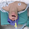
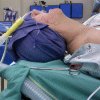
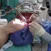
Patient in 'sniffing position' with laryngoscope suspended
-
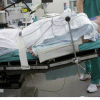
Positioning with head of bed elevated
-
Note placement of endotracheal tube to ensure it stays in the lateral sulcus. Bending the endotracheal tube to position the end inferiorly may result in movement of the tube to cross over the base of tongue.
References
Zeitels SM and Shapshay SM: Principles and Techniques of Operative Laryngoscopy: Equipment and Technology. Ch 7 in pp77 -110 in The Larynx ed Ossoff RH, Shapshay SM, Woodson GE, and Netterville JL. Lipppincott Williams & Wilkins Philadelphia 2003
Bouchayer Marc and Cornut G: Instrumental Microscopy of Benigh Lesions of the Vocal Folds. pp 143-165 Ch8 in Phonosurgery: Assesment and Surgical Managment of Voice Disorders Ed by Ford CN and Bless DM. Raven Press New York 1991
Olson GT, Moreano EH, Arcuri MR, Hoffman HT. Dental Protection During Rigid Endoscopy. Laryngoscope 105(6):662-3, 1995.
Hoffman H, Bayan S, Tokita, J, Van Daele D, Schneider: In reference to Cost-effective dental protection during rigid endoscopy Laryngoscope 2012 Oct;122(10):2362


