
return to: instrumentation - Sialendoscopy Tray (updated 3-03-2025)
Holmium laser for lithotripsy; Fixed Intermediate Sized Salivary Stones Lecture AHNS Salivary Endoscopy Course April 9 2013 (restricted); Endoscopic stone management: Intermediate Sized/Lasers/External Lecture LSU New Orleans (Hoffman) Feb 1-2 2014
see also: Sialendoscopy After Parotid Stone Fragmentation with Transillumination Composite; Pneumatic Lithotripsy; Salivary Gland Surgery Protocols; Sialolithiasis; Salivary Swelling; Sialograms and Sialography; Sialectasis
Sialendoscopy Course LSU New Orleans Lectures (Hoffman) March 21-22, 2015
Combined Open and Endoscopic Removal of Parotid Stone (sialendoscopy case example)
Parotid duct foreign body removal (sialogram, sialendoscopy)
Submandibular gland stone removal sialendoscopy case example
Submandibular Duct Foreign Body (retained salivary stent)
Sialodochoplasty (complex) for right submandibular sialadenitis with stone (sialolithiasis)
Resources for information and training in Salivary Disorders and Sialendoscopy: European Sialendoscopy Training Center
Established multidisciplinary society designed to advance the science of Salivary Disorders: European Salivary Gland Society
Koch et al (Koch 2019) through review of 66 patients treated with two different Ho:YAG laser systems identified complete fragmentation of 87 stones (parotid and submandibular) supporting Ho:YAG settings of frequency 3-6 Hz, energy level of 0.5 -1.2 J and effective power of between 3-4.8 watts.
|
Ho:YAG laser setting |
Frequency |
Power |
Energy |
|
Koch et al (Koch 2019) |
3-6 Hz (cps) |
3-4.8 Watts |
0.5-1.2Joules |
|
other recommended settings |
5 |
1.5 Watts |
Laser Settings Below: frequency 5 pulses/sec (Hz), 1.5 watts
click to see video:
GENERAL CONSIDERATIONS
- Indications
- Salivary gland swelling of unclear origin
- Obstructive sialadenitis
- Stone 73.2% / Stricture 22.6% (Turner 2009)
- Stricture 15-25% (Koch M et al 2009)
- Stricture >50% (Koch et al 2005)
- Mucus plugging
- Foreign bodies
- Evaluation for intraductal mass
- In a 2011 retrospective review of 138 pts, Koch et al. (citation "k" below) showed that Wharton's duct stenoses were associated with previous therapy and/or existing disease in 51.4% of pts. The most common conditions were surgical manipulation of the ductal system (13.8%) and allergy (26.8%). Surgical manipulation preceded stenosis distally in 17.7% of cases. Autoimmune disease was associated with 5.1% of the cases.
- Contraindications
- The only contraindication for sialendoscopy is acute sialadenitis. Although this condition is not an absolute contraindication, sialendoscopy and ductal dilation is more difficult in the setting of inflammation. In addition, use of the rigid dilator system, a semirigid endoscope, or both during an acute episode of sialadenitis increases the chance of ductal trauma and potentially fosters the spread of infection in the soft tissues of the head and neck.
PREOPERATIVE PREPARATION
- Evaluation
- History and Physical Exam
- Imaging of gland (U/S, CT, MRI, sialogram)
- CONSENT
- Describe procedure identification and dilation of duct orifice; potential problems therein
- Potential for perforation of duct, infection or other process leading to swelling with possible impact on airway (airway issue more relevant to floor of mouth swelling with submandibular gland)
- Potential for scarring of duct, inability to remove stone/deal with stricture; may lead to need for gland removal (parotidectomy or submandibular gland excision) either at the time of sialendoscopy or later. Potential for scarring to relieve symptoms but render the gland non-functional also exists (see: Salivary gland atrophy with chronic obstruction)
- Describe potential complication
- Bleeding, infection, reaction to anesthesia
- Specific: salivary fistula, lingual nerve neurosensory dysfuction (submandibular sialoendoscopy)
- Medications:
- Preoperative antibiotics (Unasyn or clindamycin)
- Preoperative oral/IV steroids
- Intraoperative administration of 3 cc of triamcinolone (Kenalog 10) through duct orifice or stent at end of case
NURSING CONSIDERATIONS
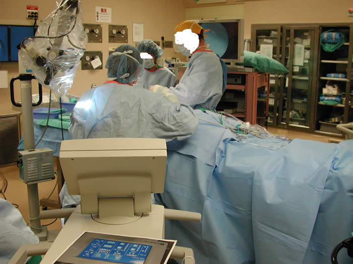
- Room Setup
- Instrumentation and Equipment
- Major Instrument Tray 1, Otolaryngology
- Major Instrument Tray 2, Otolaryngology
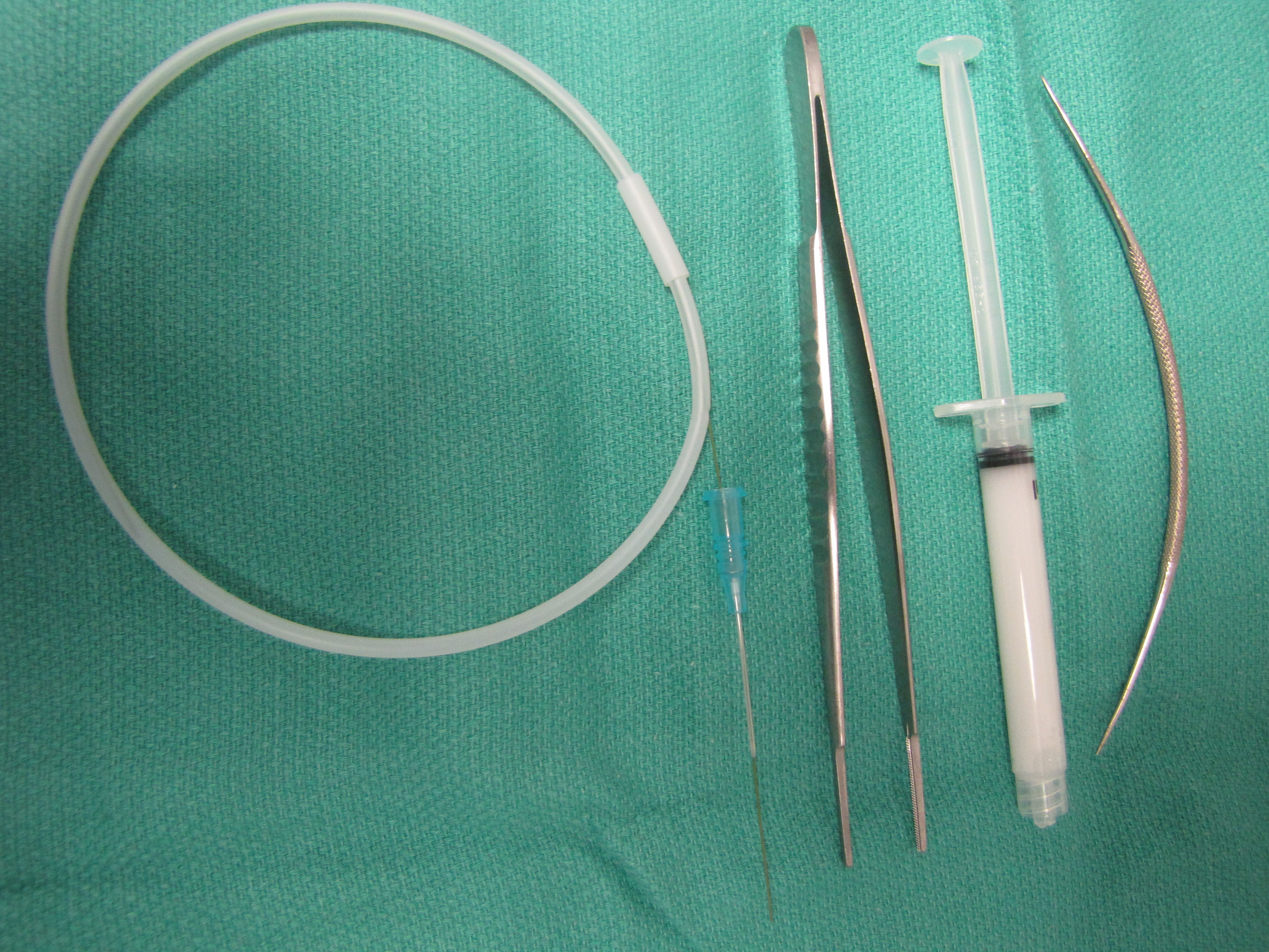
- Have available: 0.018 inch introducer (Microwire as used for angiography) which permits placement of a 22 gauge Angiocath over it by way of the Seldinger technique
- Sialendoscopy Tray
- Sialendoscopy supply basket (including baskets and balloon dilators)
- Cook Medical Salivary Access Dilator Set
- Erlengen telescopes and light cord
- 200 Micron laser fiber
- Luminis Holmium Laser Settings *** note- cases must be booked in OR room which is laser capable (with appropriate laser electrical outlet))
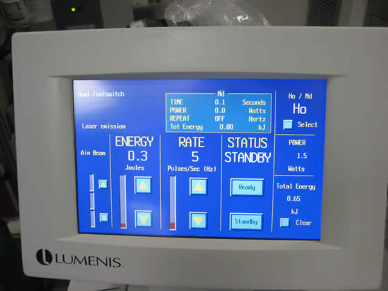
Lumenis Holmium Laser Settings
- Video unit
- Howard and Georgetown tables, Mayo stand
- Microscope with 250 lens
- Bipolar cautery unit available
- Monopolar cautery unit available
- Nerve stimulator (available for gland excision)
- Medications (specific to nursing)
- 1% lidocaine with 1:100,000 epinephrine for injection around duct (27 gauge needle) not always used - most commonly if open ductoplasty needed
- 1% lidocaine for instillation into duct at beginning of procedure (22-24 gauge angiocath)
- Kenalog 10 for instillation into duct at end of procedure (22-24 gauge needle)
- Sterile water with 20 cc syringe for infusion through irrigation port of sialendoscope
- Prep and Drape
- Oral prep with throat pack (if done under general anesthesia)
- The Spandex lip and cheek retractor and bite block (pediatric or adult) are useful for exposure once intubated. Endotracheal tube tape may make placement of the lip and cheek retractor difficult. The tube may be placed in the oral commissure contralateral to the duct of interest lateral to the retractors. The bite block should be placed with its concave surface facing laterally as this provides a convenient space for the endotracheal tube and throat pack to rest.
- Drape
- Drains and Dressings
- Special Considerations
ANESTHETIC CONSIDERATIONS
- Induction
- Antibiotics begun with placement of the IV (see Antibiotic protocol). Consider IV administration of steroids (usually decadron 8 mg IV)
- Orotracheal intubation offers adequate exposure of both Wharton's and Stensen's ducts
- Positioning
- Head of bed turned 180 degrees away from anesthesia.
- Head of bed is elevated.
OPERATIVE PROCEDURE
- Submandibular Gland
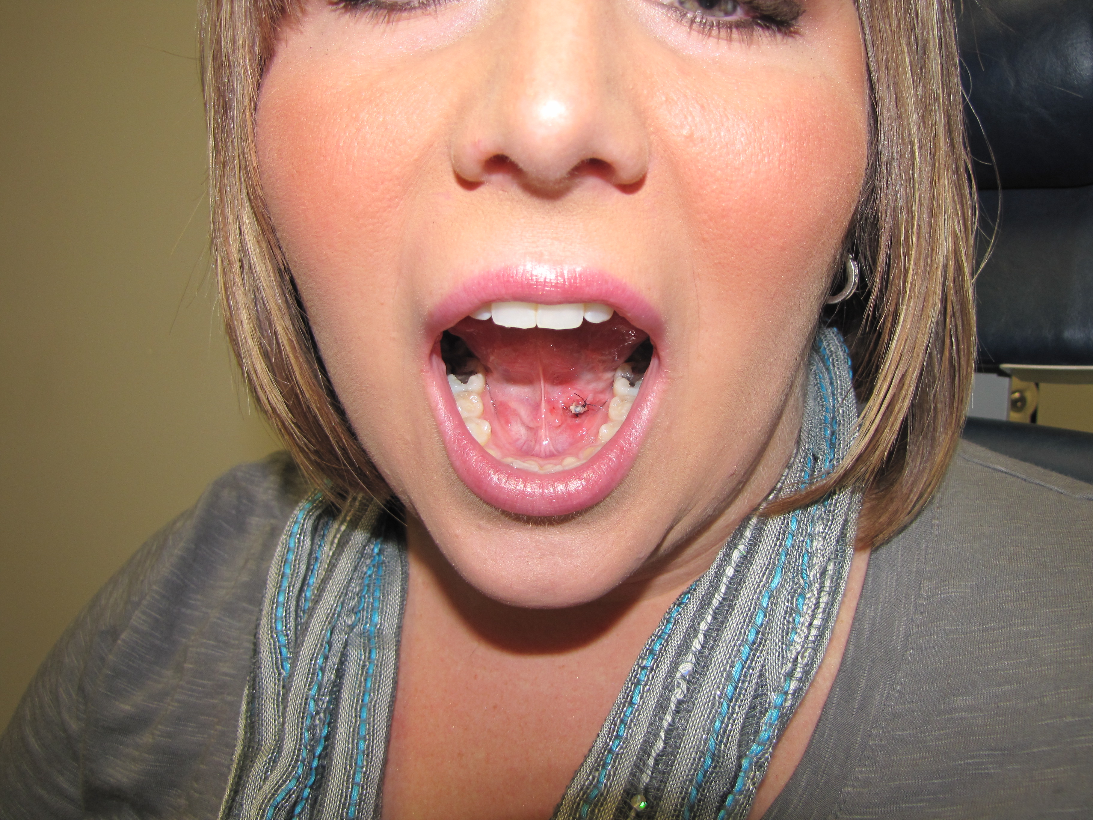
- Identify papilla (occasionally, not generally, may infiltrate papilla with 1% lidocaine with 1:100,000 epinephrine)
- Introduce salivary probes of increasing diameter
- Counter traction on tissue adjacent the papilla may be helpful
- Identification of the papilla may be facilitated by 'tipping' methylene blue onto the area of the caruncula (Luers et al reference)
- see: Salivary Cannulation and Infusion Techniques
- Introduce the dilator
- Introduce the sialendoscope; may be facilitated by introduction of a guidewire (optional)
- Parotid
- Identify papilla: Identification may be made easier by drying the buccal mucosa with compressed air, making it easier to see saliva originating from the papilla
- Cannulate duct with .018" catheter; may place 22 gauge angiocath over it, use it as a guide for the hollow bore Marchal dilators, or place it through the large (Zenk) 0.8 mm inner working channel as a guide. Papadaki et. al has described use of arterial stents for access and protection of parotid
- Infuse 1% lidocaine
- Salivary Ductoplasty with and without stent placement
- Sialolith removal techniques
- Minigrasping forceps or basket
- Mechanical fragmentation
- Intracoporeal laser fragmentation
- Combination of techniques
- See photos
- Small segment of large stone in parotid duct
- Larger segment of same stone
- Laser in place adjacent stone
- Sialodochitis after stone removal
POSTOPERATIVE CARE
- Medications
- Steroid injected intraductally. Kenalog 10, or 100 milligrams of hydrocortisone (Nahlieli)
- 7-14 days of antibiotics
Stent
- Stents may be placed in the duct and are generally left in place 3 to 6 weeks
- Appearance of stent immediately before removal three weeks after surgery:
- Postoperative management in-clinic may include steroid ductal insufflation (2-3cc of kenalog 10) see: Ultrasound Sialography (Ultrasound With Salivary Duct Infusion)
- Home care
- Sugar-free lemon drops
- Gentle massage of affected gland
- Hydration
References
Luers J-C, Vent J, and Beutner D: Methylene blue for easy and safe detection of salivary duct papilla in sialendoscopy. Otolaryngology--Head and neck Surgery (2008) 139, 466-7
Marchal F: Sialendoscopy pp 127-149(Chapter 6 in Myers E.N. and Ferris RL eds: Salivary Gland Disorders Springer, Berlin 2007)
Turner MD, Sialoendoscopy and salivary gland sparing surgery. Oral Maxilloracial Surg Clin N Am 21 (2009) 323-329
Koch M, Iro H, and Zenk J: Sialendoscopy-Based Diagnosis and Classification of Parotid Duct Stenoses. Laryngoscope, 119:1696-1703, (2009)
Koch M, Zenk J, Bozzato A, BummK, Iro H. Sialoscopy in cases of unclear swelling of the major salivary glands. Otolaryngol Head Neck surg (2005);133:863-868
Nahlieli O, Nakr LH, Nazarian Y, Turner M. Sialoendoscopy: A new approach to salivary gland obstructive pathology. JADA (2006) 137: 1394-1400.
Papadaki M, McCain JP, Kim K, Katz R, Kaban L, Troulis M. Interventional sialoendoscopy: early clinical results. J Oral Maxillofac Surg (2008) 66:954-962
Papadaki M, Kaban L, Kwolek C, Keith D, Troulis M. Arterial stents for access and protection of the parotid and submandibular ducts during sialoendoscopy. J Oral Maxillofac Surg (2007) 65:1865-1868
Brown JE. Minimally invasive techniques for the treatment of benign salivary gland obstruction. Cardiovasc Intervent Radiol (2002) 25: 345-351.
Fritsch MH: Algorithms for Treatment of Salivary Gland obstructions Without access to Extracorporeal Lithotripsy. Otolaryngologic Clinics of North America Volume 42, Issue 6, December 2009, pages 1193-1197 mfritsch@iupui.edu
Koch, M., Iro, H. et al. Diagnosis and Gland-Preserving Minimally Invasive Therapy for Wharton's Duct Stenoses.Laryngoscope 2011
Koch M, Hung SH, Su CH, Lee KS, Iro H and Mantsopoulos K: Intraducatl lithotripsy in sialolithiasis with two different Ho:YAG lasers: presetting parameters, efates. Eur Rev Med Pharmcol Sci 2019 Jul;23(13):5548-5557