


")
see: Instructions to patients submandibular salivary stones
return to: Submandibular Gland Resection
See modified operative note below identifying approach used to avoid retained stone in duct remnant. Option of retrograde sialendoscopy at end of case also possible (see reference Potash 2012).
|
|
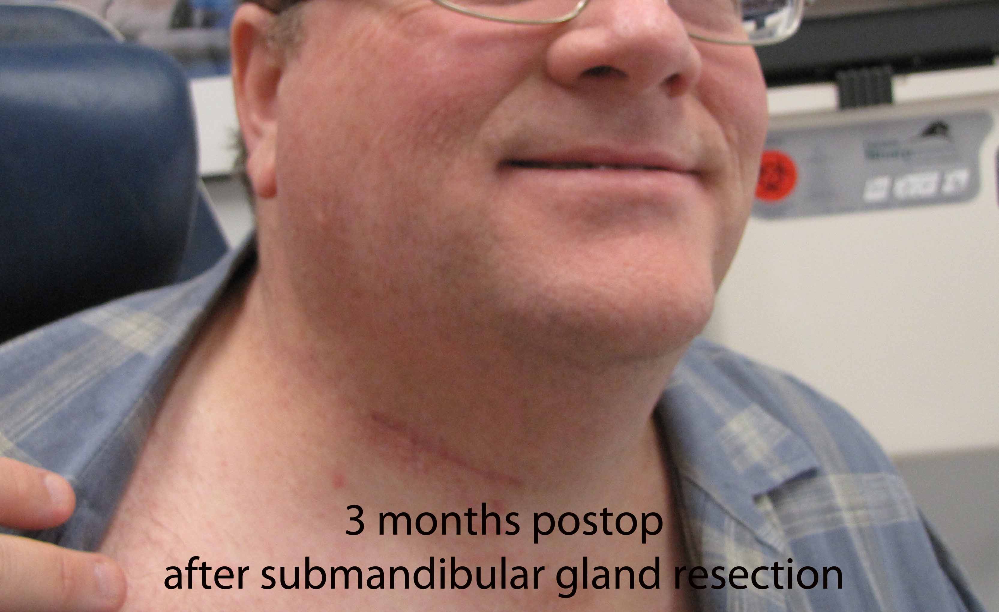
Operative Report
Indications: R submandibular gland sialithiasis and sialectasis.
Procedure Details
The patient was identified and brought into the operating room, where he was laid in a supine position and general endotracheal anesthesia induced with a GlideScope as planned without difficulty. The table was turned 180 degrees, and time out was performed. The patient was prepped and draped in sterile fashion. Operative microscope was then used to visualize the opening of the right Wharton's duct on the floor of mouth. The duct was cannulated with a 0.018 inch micro Angiocath guidewire, followed by instillation of 1% lidocaine with a 22 gauge Angiocath that was placed over the guidewire in Seldinger technique. Following that, the smallest Marshall hollow bore dilator (0000) was used to dilate the duct with tight fit, but successful dilation. The 0.8 diagnostic scope was then used to visualize the duct revealing several stones as well as a stricture and a proximal impacted stone. Further dilation of the duct permitted endoscopic basket retrieval of two small stones employing the 1.1 mm Zenk sialendoscope. Further evaluation demonstrated embedded stones adherent to wall beyond a stricture warranting submandibular gland removal.
An incision was made in the right anterolateral in a natural crease. Incision was carried down through the platysma, and the anterior border of the sternocleidomastoid was identified. Inferior border of the submandibular gland was then identified. The fascia over the submandibular gland was lifted up, with preservation of the marginal mandibular nerve. Dissection was performed around the submandibular gland capsule. Branches of the facial artery branch to the submandibular gland were isolated, sutured and divided with preservation of the integrity of the facial artery.
The mylohyoid muscle was identified and, with retraction, lingual nerve was identified and the submandibular ganglion divided after a clip was placed below the lingual nerve. Retraction sutures were placed on the submandibular duct. The gland was removed with inspection demonstrating stones in the hilum of the gland extending up to the duct. Standard antegrade sialoendoscopy was then performed with two additional stones were found in the duct which were pushed into the neck. A single final large stone was identified as adherent to the wall of the duct and could not be delivered from the duct remnant. Additional exposure through the neck required further dissection around the lingual nerve in order to successfully resect the portion of the duct under the nerve to encompass the retained stone. A hemoclip was then placed on the distal duct remnant.
The wound was irrigated with copious amounts of sterile saline. Due to retained debris in the duct remnant, as well as stenosis of the duct intraorally and intraoral edema, the distal 1 cm of duct was marsupialized in the mouth. Hemostasis was achieved intraorally with bipolar cautery and tannic acid. A 10 mm Jackson-Pratt drain was placed into the neck wound and it was closed in layers with deep 3-0 Vicryl and 4-0 nylon on the skin. The patient was then returned to Anesthesia, extubated, and taken to the Post-Anesthesia Care Unit in stable condition.
References
Potash A and Hoffman HT: Retrograde sialendoscopy: a new technique for avoiding retained ductal stones. Ann Otol Rhinol Laryngol 2012 Jan:121(1):38-43