


see also: Salivary Ultrasound
Background
1. Obesity/Diabetes
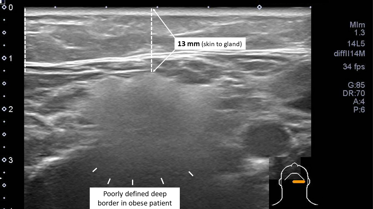
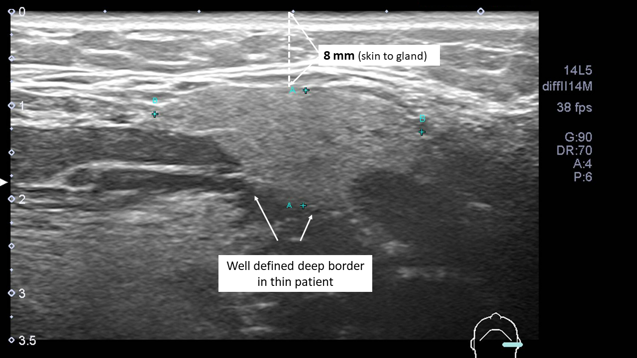
As per Badarinza (2019) the normal salivary gland has a regular contour as well as a homogeneous echotexture with intermediate echogenicity. Through their study comparing healthy patients to a group with diabetes and/or obesity - they found that those in the diabetic and/or obesity group showed increased echogenicity (hyperechoic relative to normal controls), homogeneous aspect and invisible posterior border (all p<0.001). All 18 patients in the study with enlarged parotid glands (representing sialosis subgroup) demonstrated moderate to highly increased echogencity and an invisible posterior border (but no difference in elastography).
These investigators additionally identified: "... the glandular area of salivary glands in patients with invisible posterior border was sometimes difficult to be assessed (and this can be a source of biases) and the comparison with other imaging techniques would have been useful."
Additionally "Even if the salivary glands in healthy people are described as homogeneous [19], we found a part of the glands to be rather inhomogeneous. The presence of multiple vessels in submandibular glands and the variability of longitudinal section could explain in part this inhomogeneous appearance".
In contrast: "The parenchyma of salivary glands in diabetes and/or obese patients was homogeneous in the majority of cases, probably due to the increased fatty deposition". 19 = Arya S, Pilania A, Kumar J, Talnia S. Diagnosis of bilateral parotid enlargement (Sialosis) by sonography: A case report and literature review. J Indian Acad Oral Med Radiol 2019;31:79-83
Figures below identify the hyperechoic homogeneous (smooth) echotexture with loss of deep border of the submandibular gland in an obese patient with sialosis compared to a hyperechoic (less so) homogeneous (with normal granular pattern) of normal submandibular gland.
2. Irradiation
A. External beam
Through ultrasound comparison between patients treated with irradiation (RT) for nasopharyngeal cancer (NPC) with normal controls, Cheng et al (2013) identified that the majority of the post-RT NPC patients had ill-defined borders to the submandibular glands (89%) in a statistically significant (P<0.05) greater incidence than normal controls. They postulate that high dose irradiation could destroy the normal capsule to the submandibular gland.
Chronic changes to savliary glands from external beam irradiation in the treatment of NPC have included atrophy as well as echotecture showing heterogeneity with fatty infiltration. (King 2007).
B. Radioiodine (I131)
Kim DW 2015 reported salivary ultrasound findings following I131 treatment: "post-RIA ultrasoundographic features included coarse echotexture, decreased echogenicity, lobultated maring and decreased gland size." These investigators 'hypotheiszed that post-RIA changes in the MSGs were chronic and involved profuse fatty degerenation after initial radiation-induced duct damage and consecutive acinar destruction" They reported that in some patients who underwent neck computed tomogrpahy, post-RI changes were represented by severe fatty degeration.
Previous studies (Imanimoghaddam et al. 2012; Ying et al. 2007) have reported that radiotherapy may change the echotexture, echogenicity and margins of the major salivary glands and may reduce glandular size (Kim DW 2015).
3. Chronic autoimmune sialadenitis (Sjogren's)
The lack of definition of the borders about both the parotid and submandibular glands is also a feature of diseased glands associated with chronic autoimmune sialadenitis. The finding of ‘indistinct borders’ has been included in semiquantitative grading systems for ultrasound assessment supporting the diagnosis of Sjogren's syndrome (Hocevar 2005, Milic 2010). Both contour irregularity and clarity of the posterior (deep) glandular border have been used in multiple scoring systems to contribute to a subjectively defined numerical score to identify abnormalities (Martel 2018). Although the feature of ill-defined or indistinct borders is a recognized finding, the more recent OMERACT criteria for ultrasound analysis of Sjogren’s syndrome has been truncated to a 4-point system to describe only parenchymal abnormalities and not including border assessment (Finzel 2021).
References
Kim DW. Ultrasonography Features of Normal Submandibular Parenchyma and Incidental Diffuse Submandibular Disease: A Single-Center Study. Ultrasound Q. 2019 Sep;35(3):264-268. doi: 10.1097/RUQ.0000000000000410. PMID: 30724875.
Kim DW. Ultrasonographic Features of the Major Salivary Glands after Radioactive Iodine Ablation in Patients with Papillary Thyroid Carcinoma. Ultrasound Med Biol. 2015 Oct;41(10):2640-5. doi: 10.1016/j.ultrasmedbio.2015.06.010. Epub 2015 Jul 26. PMID: 26215493.
Badarinza M, Serban O, Maghear L, Bocsa C, Micu M, Porojan MD, Chis BA, Albu A, Fodor D. Multimodal ultrasound investigation (grey scale, Doppler and 2D-SWE) of salivary and lacrimal glands in healthy people and patients with diabetes mellitus and/or obesity, with or without sialosis. Med Ultrason. 2019 Aug 31;21(3):257-264. doi: 10.11152/mu-2164. PMID: 31476205.
Kim MS, Jeong YY, Park SG, Kang NG. Age-dependent facial subcutaneous fat thickness by high-frequency medical diagnostic ultrasound system. Skin Res Technol. 2020 Sep;26(5):769-771. doi: 10.1111/srt.12858. Epub 2020 Mar 20. PMID: 32196758.
Kwon SH, Ahn GY, Lew BL, Shin JW, Na JI, Huh CH. Clinical Implication of the Regional Thickness of the Lower Facial Skin, Superficial Fat, and Superficial Musculoaponeurotic System on High-Intensity Focused Ultrasound Treatment. Dermatol Surg. 2022 May 1;48(5):527-531. doi: 10.1097/DSS.0000000000003393. Epub 2022 Jan 26. PMID: 35093961.
Taub AF. Commentary on "Clinical Implication of the Regional Thickness of the Lower Facial Skin, Superficial Fat, and Superficial Musculoaponeurotic System on High-Intensity Focused Ultrasound Treatment". Dermatol Surg. 2022 May 1;48(5):532-533. doi: 10.1097/DSS.0000000000003444. Epub 2022 Apr 5. PMID: 35384901.
Carpio Astudillo K, Montero Reyes F, Janta I, Molina Collada J, Anzola AM, Caballero Motta LR, Serrano-Benavente B, Martínez-Barrio J, Ariza Lapuente A, Rivera Redondo J, González-Fernández C, Monteagudo I, Álvaro-Gracia JM, Nieto-González JC. Salivary gland ultrasound in clinical practice: What is its real usefulness? Reumatol Clin (Engl Ed). 2023 May;19(5):260-265. doi: 10.1016/j.reumae.2022.09.002. PMID: 37147062.
Wang X, Fang W, Wei L, Li B, Cheng Y. Clinical and Sialographic Imaging Features of 131I Radiation-induced Submandibular Gland Sialadenitis. J Oral Maxillofac Surg. 2021 Feb;79(2):376-382. doi: 10.1016/j.joms.2020.07.223. Epub 2020 Aug 11. PMID: 32896506.
Cheng SC, Ying MT, Kwong DL, Wu VW. Sonographic appearance of submandibular glands in patients treated with external beam radiotherapy for nasopharyngeal carcinoma. J Clin Ultrasound. 2013 Oct;41(8):472-8. doi: 10.1002/jcu.22017. Epub 2012 Dec 1. PMID: 23203417.
King AD, Ahuja AT, Yeung DK, et al. Delayed complications of radiotherapy treatment for nasopharyngeal carcinoma: imaging findings. Clin Radiol 2007;62:195.
Nomayr A, Lell M, Sweeney R, et al. MRI appearance of radiation-induced changes of normal cervical tissues. Eur Radiol 2001;11:1807e17. (fatty infiltration)
Finzel S, Jousse-Joulin S, Costantino F, Hánová P, Hocevar A, Iagnocco A, Keen H, Naredo E, Ohrndorf S, Schmidt WA, D'Agostino MA, Terslev L, Bruyn GA. Patient-based reliability of the Outcome Measures in Rheumatology (OMERACT) ultrasound scoring system for salivary gland assessment in patients with Sjögren's syndrome. Rheumatology (Oxford). 2021 May 14;60(5):2169-2176. doi: 10.1093/rheumatology/keaa471. PMID: 33085747
Milic VD, Petrovic RR, Boricic IV, et al. Major salivary gland sonography in Sjogren’s syndrome: diagnostic value of a novel ultrasonography score (0–12) for parenchymal inhomogeneity. Scand J Rheumatol 2010;39:160–6.
Martel A, Coiffier G, Bleuzen A, Goasguen J, de Bandt M, Deligny C, Magnant J, Ferreira N, Diot E, Perdriger A, Maillot F. What is the best salivary gland ultrasonography scoring methods for the diagnosis of primary or secondary Sjögren's syndromes? Joint Bone Spine. 2019 Mar;86(2):211-217. doi: 10.1016/j.jbspin.2018.06.014. Epub 2018 Jul 24. PMID: 30053612.