click on image above, advance with cursor over border
return to: Otology - Neurotology
General
- Subtotal petrosectomy, or petrous apicectomy, involves removal of all air cells of the temporal bone, including the middle ear and mastoid in order to obtain a dry, safe ear; generally only a few air cells in the petrous apex will remain
- The otic capsule may be removed or spared
- The EAC is closed off to end in a blind sac, and the cavity is typically filled with muscle
Indications
- End-stage chronic otitis media with no possibility of hearing reconstruction, or chronic otitis media in a deaf ear
- CSF leaks
- Middle ear tumors
- Osteoradionecrosis of temporal bone
Sample Operative Note
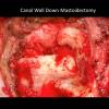
Informed consent was obtained. The patient was brought to the operating room. Anesthesia was induced. The patient was intubated and turned 180 degrees. A timeout was performed. The left ear was identified, prepped and draped in appropriate fashion, including placement of the facial nerve monitoring. A postauricular incision was planned. This was injected with 1% lidocaine with 1:100,000 epinephrine. This was taken down to temporalis fascia. An anterior-based Palva flap was raised. The spine of Henle was identified. The mastoidectomy was drilled starting with a 6-cutting burr. The tegmen, sigmoid vein, and lateral semicircular canal were identified and preserved. A 4-cutting burr was used to enter the antrum. The microscope was brought in and used for the remainder of the case. The incus was identified and removed. The entire mastoid was saucerized and mucosalized air cells were eradicated. The mastoid tip was drilled down.
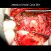
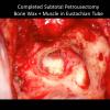
The skin was then elevated anteriorly off the posterior canal wall about 180 degrees down to the annulus. A rongeur was used to remove this posterior canal. The canal skin was transected with a 15 blade. The tympanic membrane and the medial canal skin was removed. A diamond burr was used to drill down the facial ridge. The facial nerve was identified and kept bony covered. Diamond burrs were used to drill out the mucosa of the middle ear. Temporalis muscle was harvested and was used with bone wax to plug the eustachian tube.
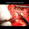
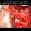
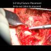
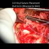
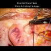
We then turned our attention to eversion of the ear canal skin. Starting inferior and medial, the tragal cartilage was identified. The anterior canal skin was freed from the tragal cartilage. The canal skin was freed from soft tissue attachments 360 degrees for full mobilization. 4-0 vicryl sutures were used to invert the skin. The first was was performed from canal skin to mucosa and then passed from mucosa to canal skin. This was done superior and inferior. A hemostat was paced through the ear canal and the Vicryls were pulled through to evert the canal skin. A total of 3 vicryls were placed. The Palva flap was rotated under the canal closure and sutured medially.
We then turned our attention to obliteration of the mastoid cavity. We then raised a temporalis flap that was pedicled anteriorly. An SCM flap that was pedicled inferiorly was raised. These muscle flaps were used to obliterate the mastoid cavity. They were approximated with 3-0 vicryl. The skin was then approximated with 3-0 vicryl. A Penrose drain was placed. The skin was closed with 3-0 nylon. Bacitracin and a mastoid dressing were applied. The patient tolerated this procedure well and was transferred to the PACU.
