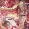
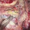
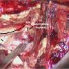
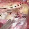
Click on image above to enlarge; advance with cursor over border
return to: Otology - Neurotology
OPERATIVE PROCEDURE
- Incision and Elevation of Skin Flaps
- Large postauricular parabolic incision. SEE FIGURE
- Raise skin and subcutaneous tissue flaps anteriorly to level of ear canal in the plane of the temporalis fascia superiorly, occipitalis fascia posteriorly, and the sternocleidomastoid fascia inferiorly.
- Preserve greater auricular nerve for grafting if necessary.
- Raise large Palva flap (from linea temporalis to mastoid tip) up to level of ear canal.
- Mastoidectomy
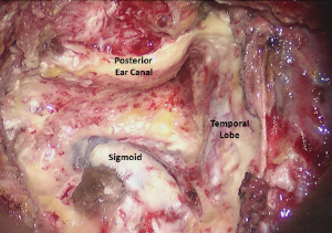
- Complete mastoidectomy with skeletonization of the dura from 2 cm posterior to the sigmoid sinus up to the bony labyrinth and down to the jugular bulb.
- The entire sigmoid to the jugular bulb should be completely decompressed.
- Must bipolar emissary veins.
- Skeletonization of the posterior bony canal and facial nerve at the second genu.
- Open the facial recess while identifying the facial nerve along its second genu.
- Complete mastoidectomy with skeletonization of the dura from 2 cm posterior to the sigmoid sinus up to the bony labyrinth and down to the jugular bulb.
- Block off Eustachian Tube
- Open epitympanum and remove incus and the head and neck of the malleus.
- Drill away the mucosa of the lateral portion of the eustachian tube.
- Plug eustachian tube with a combination of fascia, muscle, and bone wax.
- Block off middle ear with a large muscle plug.
- Labyrinthectomy
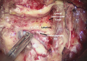
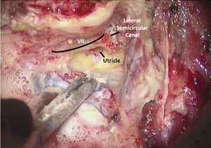
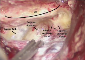
- Remove entire bony labyrinth except for the superior semicircular canal ampulla, which is the superior landmark of the internal auditory canal (IAC) and the principal landmark for the labyrinthine segment of the facial nerve (anterolateral landmark).
- Remove bone medial to labyrinth from the middle fossa dural plate to the jugular bulb.
- Spinal fluid may be seen to escape from cochlear aqueduct in smaller tumors superior to jugular bulb.
- Expose internal auditory canal
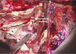
- Blueline the IAC and provide 280° of exposure medially.
- Drill away bone down to the posterior fossa dura.
- Identify Facial Nerve at Vertical Crest (Bill's Bar)
- Beware of lateral IAC superiorly due to facial nerve location.
- Superior semicircular canal ampulla is a landmark for the facial nerve that passes immediately superior and medial to it.
- Remove final pieces of bone off of IAC dura with a blunt elevator.
- Tumor Removal
- Open IAC inferiorly along the inferior vestibular nerve, with a 59-10 Beaver blade.
- Bipolar the dura overlying the cerebellum adjacent to opened IAC dura; open this dura with Beaver blade or straight microscissors as well.
- Separate the tumor from the cerebellum and place neuro cottonoids in this plane.
- Cauterize the small surface vessels; separate the larger vessels away from the tumor.
- Identify the VIIth nerve at the vertical crest and separate it from the tumor to the level of the porous acousticus.
- Bipolar the tumor pseudocapsule, away from the facial nerve, and debulk the tumor from within using either cup forceps and suction or the CUSA dissector .
- Identify the facial nerve medial to the porous. Usually it is directly anterior, but it may be superiorly or inferiorly displaced.
- Identify the VIIth and VIIIth nerves at the brainstem.
- Perform further debulking as necessary.
- Separate the VIIIth nerve and tumor from the VIIth nerve at the brainstem up to the porous (work from both ends of the facial nerve to connect them).
- Separate the tumor and associated VIIIth nerve from the brainstem and cerebellum.
- Closure
- Irrigate and check hemostasis intracranially
- Place Oxycel or Avitene along the brainstem vasculature
- Dural repair
- 4-0 Neurolon suture should be used to reapproximate the dura, or to suture in a dural graft
- First choice = temporalis fascia
- Alternatives: Durapair™ Duragen™, rectus fascia.
- Fascia should be draped over facial ridge
- Tisseal™ is optional to further seal dural area
- 4-0 Neurolon suture should be used to reapproximate the dura, or to suture in a dural graft
- Mastoid/cranial defect
- Use left lower quadrant abdominal fat, cut into strips, to fill the mastoid defect
- ¼ inch Penrose drain left in abdominal wound
- Two-layer closure of abdomen
- Use left lower quadrant abdominal fat, cut into strips, to fill the mastoid defect
- Skin/subcutaneous tissues
- Close the Palva flap over the abdominal fat and fascia with water-tight interrupted 3-0 vicryl sutures.
- Close the subcutaneous tissues with interrupted 3-0 vicryl sutures .
- Close skin with running/locking 3-0 nylon.
- Bulky mastoid-type dressing with adequate pressure to prevent spinal fluid subgaleal effusion from developing.
