Return to: Buccal Mucosa Graft for Urethral Reconstruction or Case Example of Buccal Fat Flap Palate Reconstruction
Buccal Mucosa Anatomy
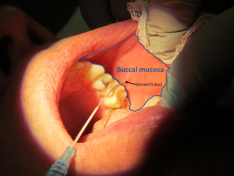
The buccal mucosa is bordered vertically by the maxillary and mandibular vestibular folds, whereas its anterior and posterior borders are formed by the outer commissure of the lips and the anterior tonsillar pillar, respectively. The buccal mucosa is primarily innervated by the long buccal nerve (CN V3), and by the anterior, middle, and posterior superior alveolar nerves of the second division of the trigeminal nerve (CN V2). Additionally there is limited sensory innervation from the facial nerve. The blood supply of the buccal mucosa originates primarily from:
- The buccal artery---a branch of the maxillary artery
- The anterior superior alveolar artery of the infraorbital artery---a branch of the third part of the maxillary artery
- The middle and posterior superior alveolar arteries---branches of the maxillary artery
- Accessory vessels from the transverse facial artery---a branch of the superficial temporal artery
The harvesting surgeon must continually be aware of the parotid (Stensen's) duct. This duct originates from the parotid gland, travels across the body of the masseter muscle, turns medially at the anterior border of the masseter, crosses the buccal fat pad, pierces the buccinator muscle, and finally terminates at its orifice. This orifice is clinically visible by a papilla located on the mucosa of the inner aspect of the cheek adjacent to the maxillary second molar.
The anatomic buccal space located lateral to the buccinator muscle consists of: adipose tissue (buccal fat pad), Stensen’s duct, the facial artery and vein, lymphatic vessels, minor salivary glands, and branches of CN 7 and CN 9. The surgeon must be aware of these structures underlying buccinator muscle. However, they should not be exposed in the process of harvesting the buccal mucosal graft which is dissected lateral to the buccinator muscle. Failure to leave the buccinator muscle intact could result in poor wound closure at the donor site and dysfunction of the buccinator muscle, which serves as a muscle of facial expression.
The mucous membrane lining the cheek is reflected above and below upon the gums is continuous behind with the lining membrane of the soft palate. Opposite the second molar tooth of the maxilla is a papilla, on the summit of which is the aperture of the parotid duct. (Gray's Anatomy 1918)

Masticator Space
Located:
- inferior to the temporal space
- anterolateral to the parapharyngeal space
- the masticator space lies lateral to the infratemporal fossa
Contains:
- masseter muscle
- the external (lateral) and internal (medial) pterygoid muscles
- the posterior body and the ramus of the mandible
- the inferior alveolar vessels and nerves
- the insertion of the temporalis muscles
The space is traversed by the mandibular nerve and internal maxillary vessels.
Masticator space is formed superficial layer of the deep fascial surrounding loose connective tissue and fat pad along with the above structures.
The masticator space is further subdivided into the following compartments:
- Masseteric
- Pterygoid
- Superficial temporal
- Deep temporal
Each of these subdivisions of the masticator space require directed approach to address masses or infection
- The most common source of infection is dental in origin, most commonly the third molar.
Due to close proximity of the muscles of mastication, patients with infections often have severe trismus as well as tenderness along the mandible
- Suspicion of metastasis/malignancy should lead to clinical evaluation of V3, which exits through foramen ovale into the masticator space.
Surgical approaches may be made either via an intraoral incision along retromolar trigone area or via an extraoral approach along the inferior mandible.
References
Henry Gray (1821--1865). Anatomy of the Human Body. 1918
Arlen AM, Powell CR, Hoffman HT, Kreder KJ. Buccal mucosal graft urethroplasty in the treatment of urethral strictures: experience using the two-surgeon technique. ScientificWorldJournal. 2010 Jan 8;10:74-9. doi: 10.1100/tsw.2010.16. PMID: 20062954; PMCID: PMC5763984.
Chughtai S, Chughtai KA, Montoya S, Bhatt AA. Radiographic review of anatomy and pathology of the masticator space: what the emergency radiologist needs to know [published correction appears in Emerg Radiol. 2020 Mar 14;:]. Emerg Radiol. 2020;27(3):329-339. doi:10.1007/s10140-020-01756-7