The Cranial Nerves

I. Olfactory Nerve

- Cell bodies located in the olfactory mucosa overlying the superior nasal concha and superior septum
- Axons pass through the cribiform plate of the ethmoid bone
- Forms olfactory bulb that connects to the brain via the olfactory tract
- Function: Smell
- Otolaryngic Significance:
- Anosmia: DDx: obstruction (tumor, sinus disease, deviated septum, nasal polyps), medications, tobacco, aging, parkinsons, trauma
II. Optic Nerve


- Cell bodies located in the ganglionic layer of the retina
- Course: Optic nerve leaves orbit via optic canal of sphenoid bone, runs postero-medially towards the optic chiasm
- Function: Vision
- Otolaryngic Significance:
- Orbital Hematoma - occurs secondary to trauma, sinus surgery, neurosurgery and ophthalmic surgery can cause optic neuritis and decreased visual acuity
- Sellar lesions - lesions in the sella turcica (pituitary adenoma, craniopharyngioma) are in close proximity to optic chiasm and can result in mass effect on optic nerve and classically causes bitemporal hemianopsia
- See Radiographic sellar anatomy, Radiographic anatomy of the orbit, Transsphenoidal Approach to Pituitary Gland
- Orbital cellulitis/abscess, cavernous sinus thrombosis - infectious process involving soft tissues of the orbit posterior to the orbital septum or cavernous sinus can result in inflammation of or mass effect on the optic nerve resulting in acute visual acuity loss
III. Oculomotor Nerve
- Course: Arises from brain stem, passes anteriorly through the cavernous sinus, leaves cranium via superior orbital fissure, enters orbit and divides into superior and inferior divisions
- Function:
- Skeletal motor supply
- Superior division: superior rectus and levator palpebrae superioris muscles
- Inferior division: medial rectus, inferior rectus and inferior oblique muscles
- Parasympathetic
- Oculomotor carries parasympathetic innervation from the Edinger-Westphal nucleus to the ciliary ganglion which supplies sphincter pupillae (pupil constriction) and ciliary muscle (accommodation)
- Sympathetic
- Oculomotor carries fibers from the peri-carotid sympathetic plexus of nerves which supply superior tarsal (Mueller's) muscles
- Skeletal motor supply
- Otolaryngic significance:
- Trauma:
- Lateral blunt head trauma may result in epidural hematoma secondary to middle meningeal artery rupture. Herniation of the brain can causes direct pressure on the oculomotor nerve
- Orbital blowout fracture may cause entrapment of inferior or medial rectus muscles mimicking oculomotor injury
- Cavernous sinus pathology (thrombosis, abscess, tumor, AVM)
- Horner's Syndrome: disruption of sympathetic cervical chain causes ptosis, anhidrosis, and miosis
- Classically associated with Pancoast tumor, but can result from many pathologies of the head and neck
- Thyroid carcinoma, goiter, thymoma
- Iatrogenic (neck dissection, thyroidectomy, parathyroidectomy)
- Trauma (blunt or penetrating)
- Cavernous sinus thrombosis (post-ganglionic = no anhidrosis)
- Classically associated with Pancoast tumor, but can result from many pathologies of the head and neck
- Trauma:
IV. Trochlear Nerve
- Course: Arises from the brain stem, courses in the subarachnoid space superoanteriorly, pierces the dura to enter the cavernous sinus, enters the orbit via the superior orbital fissure.
- Function: Motor supply to the superior oblique muscle (up gaze and superior torsion)

V. Trigeminal Nerve
- Course: Sensory and Motor roots arise from brainstem and form semilunar ganglion in Meckel's cave, from there, 3 divisions arise: ophthalmic branch, maxillary branch and mandibular branch (commonly called V1, V2 and V3 respectively)
- Otolaryngic significance:
- A thorough understanding of the trigeminal nerve anatomy may be utilized for very effective local anesthesic blocks used in many procedures of the head and neck including nasal fracture reduction, laceration repair, excision of facial lesions, local flap repair, septal hematoma drainage, dental procedures, and intraoral procedures.
- Trigeminal neuralgia
- Infraorbital nerve FESS
- Blowout fracture
V1 - Ophthalmic Nerve
From Meckel's cave, it pierces the dura to enter the cavernous sinus, leaves the cranium via the superior orbital fissure and divides into nasociliary, frontal and lacrimal nerves.
- Nasociliary nerve:
- Course: enters the orbit between the two heads of the lateral rectus muscles and between the superior and inferior rami of the oculomotor nerve. Runs medially through the orbit and continues anteriorly along the medial wall of the orbit.
- Branches: (PLICA mneumonic)
- Communicating branch to ciliary ganglion
- Long Ciliary Nerve - provides sensation to cornea/sclera, carries sympathetic fibers from the peri-carotid plexus of nerves to the ciliary body and dilator pupillae muscle
- Infratrochlear nerve - continues anteriorly through the orbit towards the medial commisure, it pierces the medial canthal ligament and supplies sensory innervation to the upper eyelids, bridge of the nose and conjunctiva.
- Posterior ethmoidal nerve - passes through posterior ethmoidal foramen and supplies sensory innervation to the mucosa of the sphenoid sinus, posterior ethmoid sinuses and posterior lateral nasal cavity
- Anterior ethmoidal nerve - passes through anterior ethmoidal foramen, provides sensory innervation to anterior ethmoid sinus mucosa, continues back into the cranium via the cribiform plate and returns to the nasal cavity through the nasal slit to provide sensory innervation to the superior nasal septum
- Frontal nerve:
- Course: enters orbit as superior most structure of the orbit between the levator palpebrae superioris and the periosteum
- Branches:
- Supratrochlear nerve - medial branch, exits the orbit medial to the pulley and courses superiorly along the cranium deep to the frontalis to provide sensory innervation to the scalp of the anterior head near the midline
- Supraorbital nerve - the lateral branch, exits the orbit via the supraorbital foramen and courses superiorly deep to the frontalis innervate the scalp over the frontal and parietal areas, supplies mucosa of frontal sinus
- Lacrimal nerve:
- Course: enters orbit as lateral most structure and courses along lateral orbital wall with lateral rectus muscle
- Function: Sensory innervation to lateral upper eyelid, parasympathetic innervation to lacrimal gland (from pterygopalatine ganglion via communicating branch between zygomatic nerve (see below) and V1.


V2 - Maxillary Nerve
From Meckel's cave, the nerve pierce the dura to enter the cavernous sinus and leaves the cranium via the foramen rotundum into the pterygopalatine fossa and gives off the infraorbital nerve, zygomatic nerve, nasopalatine nerve, superior alveolar nerves, palatine nerves, and pharyngeal nerve.
- Infraorbital nerve:
- Course: from pterygopalatine fossa, the maxillary nerve gives off the infraorbital nerve to the infraorbital canal in the floor of the orbit/roof of the maxillary sinus and exits onto the face via the infraorbital foramen
- Function: Sensory innervation to the inferior eyelid, superior lip, lateral nose and vestibule
- Zygomatic nerve:
- Course: from the pterygopalatine fossa, the maxillary nerve gives off the zygomatic nerve which passes through the inferior orbital fissure into the orbit, the nerve exits the orbit laterally through the zygomaticofacial foramen to the face. A branch of the zygomatic nerve also carries parasympathetic fibers from the pterygopalatine ganglion to the lacrimal nerve of V1 providing parasympathetic input to the lacrimal gland.
- Function: Parasympathetic motor innervation of the lacrimal gland, sensory innervation of the lateral forehead and the cheek
- Nasopalatine nerve:
- Course: from the pterygopalatine fossa, the nasopalatine nerve enters the nasal cavity via the sphenopalatine foramen. The nerve crosses the posterior roof of the nasal cavity just inferior to the sphenoid os to reach the lower 1/3 of the septum. It runs anteriorly along the septum between the mucosa and periosteum towards the incisive canal where it enters the oral cavity and innervates the anterior hard palate.
- Funtion somatic sensory innervation of the nasal septal and anterior palate mucosa. "The Pizza Burn Nerve"
- Superior alveolar nerve:
- Course: from the pterygopalatine fossa, the maxillary nerve gives off the superior alveolar nerve which quickly divides into 3 smaller nerves.
- Posterior superior alveolar nerve exits the pterygomaxillary fissure and runs over the maxillary tuberosity. The nerve arborizes and multiple branches enter several small foramina in the maxilla to reach the posterior maxillary dental roots. Other branches enter the oral cavity to innervate the posterior maxillary gingivae.
- Middle superior alveolar nerve branches from the infraorbital nerve into the maxillary sinus and runs along the wall of the maxillary sinus (innervating the maxillary sinus mucosa) to reach the maxillary dental roots of the pre-molars.
- Anterior superior alveolar nerve branches from the infraorbital nerve just before the infraorbital foramen. The nerve descends along the anterior wall of the maxillary sinus in a canal to reach the dental root of the maxillary canine and incisors.
- Function: sensory innervation of the maxillary dentition and contribution to sensory innervation of paranasal sinuses.
- Course: from the pterygopalatine fossa, the maxillary nerve gives off the superior alveolar nerve which quickly divides into 3 smaller nerves.
VII: Facial Nerve
(See more facial nerve anatomy) (See even more facial nerve anatomy)
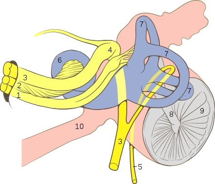
- Course: the facial nerve forms at the lateral surface of the brainstem at the pontomedullary junction along with CN VIII. Both CN VII and VIII course laterally to the internal auditory meatus. (see the image to the right for the relationship of the nerves in the IAC). From the IAC, the facial nerve takes a very anatomically complex and tortuous course through the facial canal of the temporal bone. This very detailed anatomy is for the most part, low yield for medical students, but several key points will be discussed.
- Nervus intermedius: along with the true motor facial nerve, the intermedius nerve follows the facial nerve and carries parasympathetic pre-ganglionic fibers and special sensory efferent fibers. The distribution of these will be discussed below. From this point on, the facial and intermedius nerves will be referred together as the facial nerve or CN VII.
- Labyrinthine part: From the IAC, the facial nerve courses laterally through petrous temporal bone and passes between the cochlea and the vestibular apparatus.
- Anterior genu: within the petrous temporal bone, the facial nerve takes a sharp turn to take a posteroinferior course. At this turn, a swelling in the nerve forms wherein, special sensory efferents have their cell bodies in the geniculate ganglion.
- Greater petrosal nerve: From the geniculate ganglion, a pre-ganglionic parasympathetic afferent branch exits the petrous bone into the cranium forming the greater petrosal nerve. This nerve enters the vidian (also called pterygoid) canal to enter the pterygopalatine fossa. The fibers synapse with post-ganglionic parasympathetics which are distributed along the branches of V2 to provide parasympathetic motor to the mucosa of the paranasal sinuses, nasal cavity and superior oral cavity. A communicating branch to V1 provides parasympathetic motor innervation to the lacrimal gland.
- Tympanic part, after the genu, the facial nerve enters the tympanic cavity and runs posteriorly medial to the ossicles. The facial nerve exits the tympanic cavity through the pyramidal process where it turns and forms the posterior genu.
- Mastoid part: The facial nerve courses inferiorly from the posterior genu to the stylomastoid foramen where it exits the cranium. The mastoid part gives off chorda tympani.
- Chorda tympani: the chorda tympani is a branch of the mastoid part of the facial nerve. The chorda courses posterior to anterior back into the tympanic cavity where it runs essentially along the medial surface of the superior tympanic membrane. The chorda exits the tympanic cavity via the petrotympanic fissure where it enters the infra temporal fossa and joins the lingual nerve. The chorda carries pre-ganglionic parasympathetics to the submandibular ganglion and special sensory (taste) efferents to the anterior 2/3 of the tongue. The submandibular ganglion supplies parasympathetic motor innervation to the submandibular, sublingual and minor salivary glands.
Intracranial Facial Nerve Anatomy

Vestibular nerve
Cochlear nerve
Intermediofacial nerve
Anterior genu/Geniculate ganglion
Chorda tympani
Cochlea
Vestibular apparatus
Malleus
TM
- Facial part: the facial nerve exits the stylomastoid foramen as one main trunk called the pes anserinus. Immediately, the posterior auricular nerve branches posteriorly. The nerve then passes through the parotid gland (which it does not innervate) in a deep to superficial course. Within the parotid, the facial nerve branches extensively. The first 2 divisions of pes anserinus are typically a temporofacial trunk and a cervicofacial trunk. From here a plexus of nervous interconnections forms and terminates in 5 distributions: Temporal, Zygomatic, Buccal, Mandibular and Cervical (To Zanzibar By Motor Car).
- Motor innervation: The facial nerve supplies motor innervation to all muscles of facial expression, the posterior belly of the digastric muscle and the stapedius muscle.
Extracranial Facial Nerve Anatomy

Facial Nerve Schematic
- Function:
- Somatic motor: Motor to muscles of facial expression, stapedius and posterior belly of digastric
- Parasympathetic motor:
- Paranasal sinuses, nasal cavity, oral cavity mucosa, minor salivary glands and lacrimal gland via greater petrosal nerve --> pterygopalatine ganglion
- Submandibular and sublingual glands as well as minor salivary glands and oral mucosa of inferior oral cavity via chorda tympani --> submandibular ganglion
- Special sensory: Taste to anterior 2/3 of tongue via chorda tympani to lingual nerve.
- Otolaryngic significance:
- Bell's Palsy
- Trauma
- Lacerations can sever facial nerve branches and require neurorrhaphy
- Temporal bone fractures can cause facial nerve swelling or injury
- Parotid neoplasms: parotidectomies require meticulous dissection of the parotid gland off of branches of the facial nerve
- Neck dissection: An early step of neck dissection is the identification of the marginal mandibular nerve deep to the superior sub-plastysmal flap. Injury to the marginal mandibular nerve is a very morbid complication resulting in oral incompetence.
- Prep for the OR:
- In what plane will we find the facial nerve?
Understand that the distal branches of the facial nerve enter the muscles of facial expression on the deep surfaces. The facial nerve runs in the deep layer of the SMAS (superficial musculoaponeurotic system)
What are some ways to identify the facial nerve?
Be ready for this question!! There are several classic answers:
- 1 cm deep, 1 cm anterior and 1 cm inferior to the inferior point of the external canal cartilage (also known as the "Tragal Pointer"
- 1 cm deep to the attachment of the posterior belly of digastric to the digastric groove of the mastoid
- Follow the plane of the tympanomastoid suture, the facial nerve will be in this plane 0.5 to 1 cm distal to the fissure
- In some unusual circumstances, a mastoidectomy to identify the mastoid portion of the facial nerve can be done and the nerve can be followed peripherally (this is rare)
- Identifying a peripheral branch of VII allows for retrograde dissection. Some of the landmarks for these nerve branches are:
- A branch of VII typically crosses superficially over the facial vein