see: Acute Facial Paralysis Evaluation
General
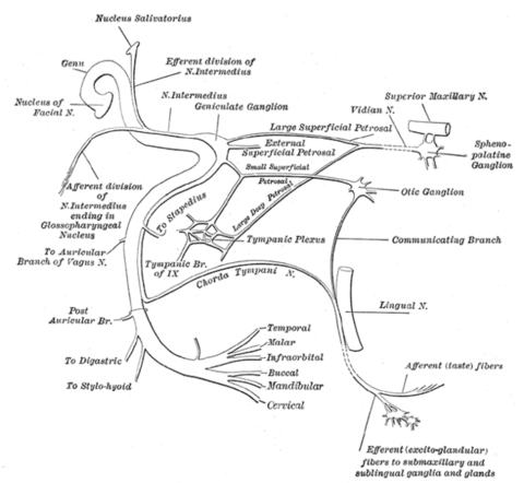
Cranial nerve seven (CN VII) is responsible for both efferent and afferent modalities in the head and neck including:
- Branchial motor fibers that innervate:
- muscles of "facial expression"
- stylohyoid muscle
- posterior belly of digastric
- stapedius
- occipitofrontalis
- Special sensory fibers for taste from the anterior 2/3 of tongue
- General sensation from the external auditory canal and auricle
- Preganglionic parasympathetic fibers innervating the submandibular, sublingual, lacrimal glands as well as the mucous membranes of the nose, palate, and oropharynx

Extracranial Course
- Soon after exiting the stylomastoid foramen, the facial nerve gives rise to three motor nerves:
- Posterior auricular nerve (occipitofrontalis muscle)
- Nerve to the posterior belly of the digastric muscle (raises hyoid bone)
- Nerve to the stylohyoid muscle (raises hyoid bone)
- The remaining motor fibers of the facial nerve continue anteriorly/inferiorly and enter the parotid gland. Within the parotid gland, the facial nerve divides into five branches:
- Temporal
- Zygomatic
- Buccal
- Marginal mandibular
- Cervical
- A popular mnemonic to remember these intra-parotid divisions is:
- “To Zanzabar By Motor Car”
- These branches of the facial nerve are responsible for providing motor innervation to the muscles of “facial expression,” which are frequently tested during physical examination.
- It is important to note that while the facial nerve branches within the parotid gland, it does not provide autonomic innervation to the gland
- This is supplied by the auriculotemporal division of V3.
- It is essential to identify and preserve these five branches when performing a parotidectomy
See: Parotidectomy with Facial Nerve Dissection
Branchial Motor Innervation
- The largest component of CN VII provides efferent motor innervation to the following muscles:
- Muscles of “facial expression”
- Stylohyoid m. (raises hyoid bone)
- Posterior belly of digastric m. (raises hyoid bone)
- Stapedius m. (dampens vibrations of the stapes on the oval window)
- Occipitofrontalis m. (intrinsic muscle of the scalp)
- The muscles innervated by CN VII are known as “branchial muscles” as they are derived from embryonic structures of the 2nd pharyngeal arch (hyoid arch).
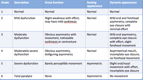
- CN VII is often tested during physical examination and its function can be reported using the House-Brackmann Scale (Hint: 1 is normal, 6 is completely non-functional).
Parasympathetic Innervation
- CN VII provides preganglionic parasympathetic innervation to:
- Submandibular glands
- Sublingual glands
- Lacrimal glands
- Mucous membranes of the nose, palate, and pharynx
- It is important to note that all of the postganglionic parasympathetic nerve fibers from CN VII are actually carried to their ultimate targets via divisions of CN V.
- Parasympathetic innervation in the head and neck promotes the production of mucous, tears, and saliva and is counter regulated by sympathetic innervation.
Anatomic Pathway of Parasympathetics
- The parasympathetic fibers of CN VII originate in the superior salivary nucleus of the pons and leave the cerebellopontine angle as the nervus intermedius (of Wrisberg).
- The nervus intermedius enters the internal acoustic meatus (IAC).
- The parasympathetic fibers then pass through the geniculate ganglion (without synapsing) and join one of two divisions of CN VII:
- Greater petrosal nerve (parasympathetics only)
- Chorda tympani (taste to anterior 2/3 tongue as well as parasympathetics)
Greater Petrosal Nerve
- The parasympathetic preganglionic fibers that join the greater petrosal nerve travel anteromedialy and enter the middle cranial fossa, passing over the foramen lacerum.
- These fibers then enter the pterygoid canal of the temporal bone (along with sympathetic fibers of the deep petrosal nerve) and form the pterygoid nerve.
- The pterygoid nerve travels through the pterygoid canal until it reaches pterygopalatine fossa.
- Here, the preganglionic parasympathetic nerve fibers synapse in the aptly named pterygopalatine ganglion.
- The postganglionic parasympathetic fibers then join branches of the V2 division of CN V and provide innervation to:
- Lacrimal glands
- Mucous membranes of the nose, palate, and oropharynx
Chorda Tympani
- The parasympathetic preganglionic fibers that join the chorda tympani enter the temporal bone and cross the tympanic membrane.
- They then exit the cranium via the petrotympanic fissure and join the lingual branch of cranial nerve V3 in the infratemporal fossa.
- The lingual nerve travels anteroinferiorly through the floor of the mouth, where the preganglionic fibers synapse in the submandibular ganglion.
- The postganglionic fibers then travel a short distance to innervate their two final targets:
- Submandibular salivary glands
- Sublingual salivary glands
see: Botox Injection for Freys Syndrome
Chorda Tympani:
- The chorda tympani provides taste sensation from the anterior 2/3 tongue and also carries preganglionic parasympathetic nerve fibers that innervate the submandibular and sublingual salivary glands.
- The nerve fibers of the chorda tympani arise from 2 distinct nuclei in the pons:
- Superior salivary nucleus:
- Origin of parasympathetic fibers
- Nucleus tractus solitarius (NTS):
- Destination of afferent taste fibers
- Superior salivary nucleus:
- The fibers from the superior salivary nucleus and the NTS converge as they leave the cerebellopontine angle and enter the internal acoustic meatus (IAC).
- They then travel through the facial canal where only the taste fibers synapse in the geniculate ganglion.
- At this point, the newly arisen postsynaptic afferent taste fibers and the presynaptic parasympathetic fibers travel with the facial nerve through the facial canal.
- Before the facial nerve exits the cranium via the stylomastoid foramen, it gives off the chorda tympani.
- The chorda tympani travels superiolaterally and enters the middle ear, arches across pars flaccida medial to the superior portion of the handle of malleus, and traverses above the insertion of tensor tympani.
- The chorda tympani exits the cranium via the petrotympanic fissure and enters the infratemporal fossa.
Examination
- The facial nerve is responsible for the voluntary movements of the "muscles of facial expression."
- The 5 divisions of CN VII innervate different regions of the face:
- Orbital group: the orbicularis oculi is the only muscle that closes the eye.
- Paralysis leads to ectropion (the lower eyelid turns outward, exposing the inferior globe)
- This may lead to drying and ulceration of the cornea if left unprotected.
- Paralysis leads to ectropion (the lower eyelid turns outward, exposing the inferior globe)
- Nasal group: nasalis, procerus, depressor septi nasi
- Oral group: orbicularis oris, buccinator, zygomaticus major/minor
- Paralysis causes difficulty in feeding or sialorrhea (drooling)
- Orbital group: the orbicularis oculi is the only muscle that closes the eye.
Table adapted from House and Brackmann (1985)

References
Scott Hirsch,Geoffrey Ferril,Adam M. Terella. Chapter 66 Facial Reanimation. ENT Secrets. Elsevier 2023
Warner MJ, Hutchison J, Varacallo M. Bell Palsy. 2023 Aug 17. In: StatPearls [Internet]. Treasure Island (FL): StatPearls Publishing; 2023 Jan–. PMID: 29493915.
House JW, Brackmann DE. Facial nerve grading system. Otolaryngol Head Neck Surg. 1985 Apr;93(2):146-7. doi: 10.1177/019459988509300202. PMID: 3921901.