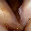
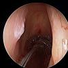
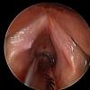
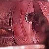
Click on image to enlarge; advance with cursor over right mid border
return to: Laryngeal leukoplakia white plaques on vocal cords
see also: Esophagoscopy with narrow band imaging (NBI) for Reflux Esophagitis
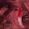
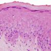
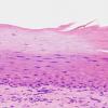
Pathology showed: Focal mild dysplasia at anterolateral margin with associated acanthosis, hyperkeratosis, and acute and chronic inflammation.
Biopsy of irregular GE junction showed Barrett's metaplasia without dysplasia.
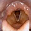
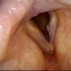
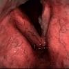
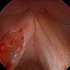
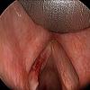
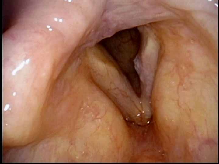
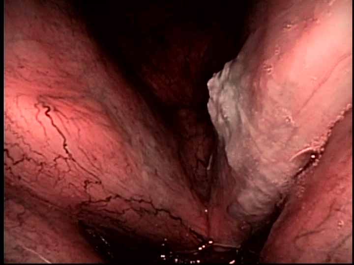
Preoperative Exam in clinic with transnasal high definition videostroboscopy with narrow band imaging |
|||
|---|---|---|---|
 |
 |
 |
 |
|
High definition standard light |
High definition narrow band imaging |
High definition with narrow band imaging (NBI) |
|
 |
|||
|
High definition standard light |
|||
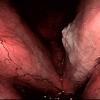
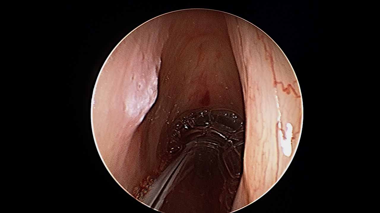
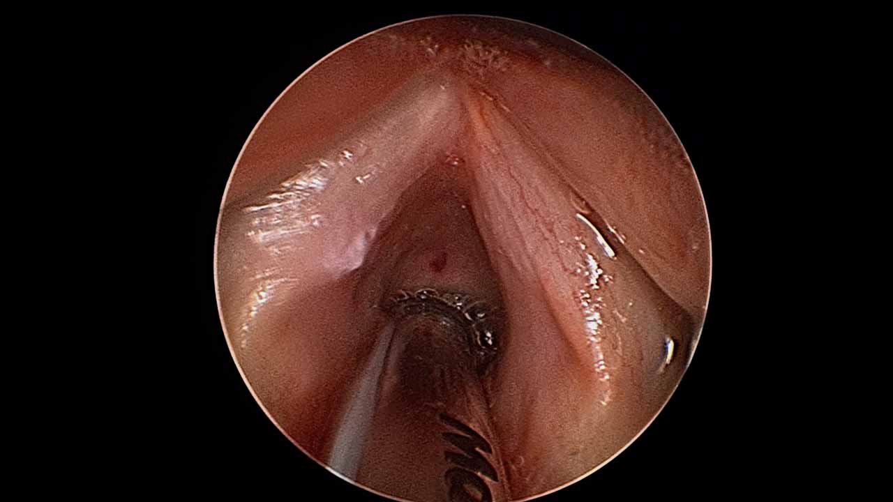
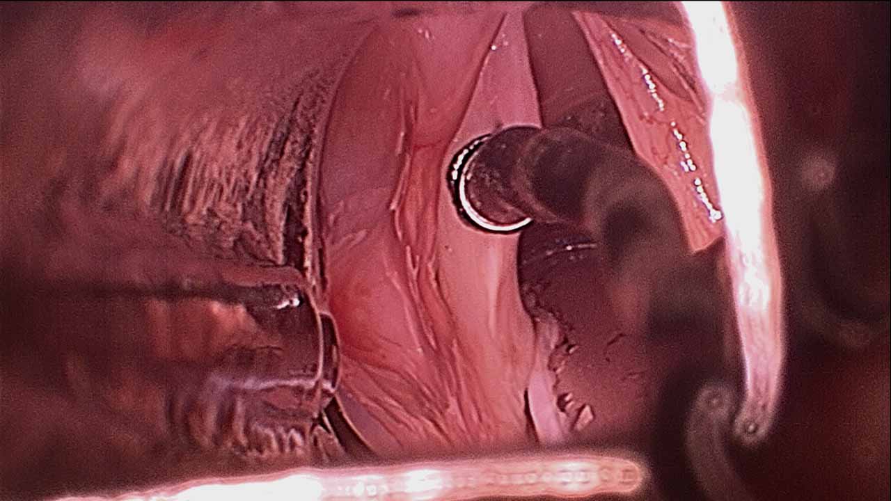
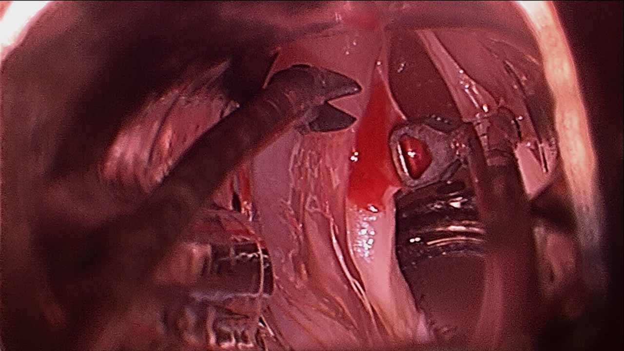
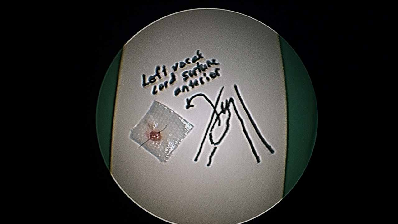
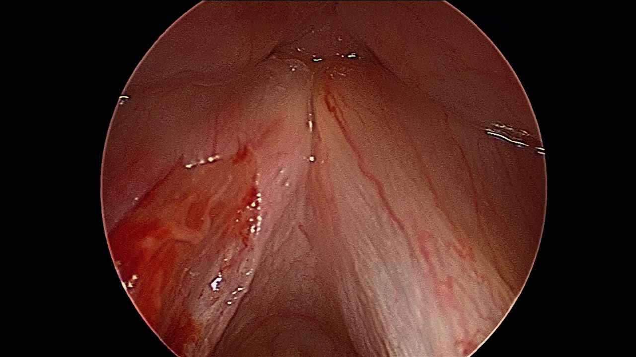
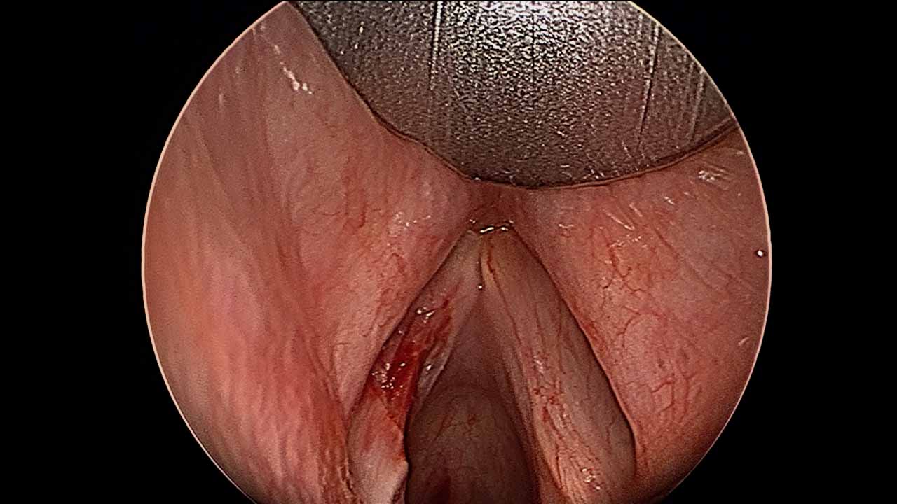
Intraoperative resection left vocal fold leukoplakia |
|||
|---|---|---|---|
 |
 |
 |
 |
 |
 |
 |
|
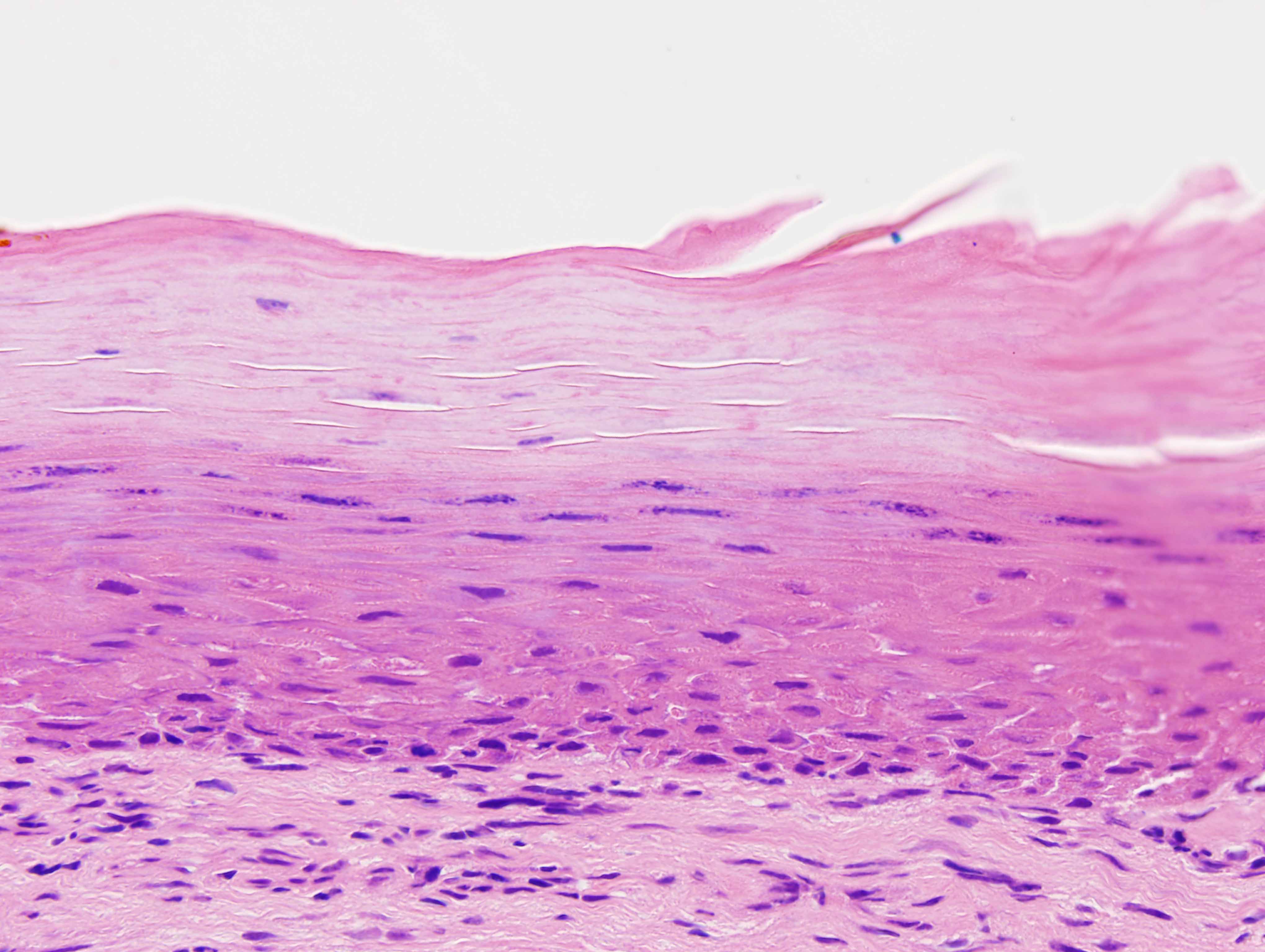
Histology of left vocal cord resection |
|
|---|---|
 |
 |
|
This mucosa shows budding along the mucosa/submucosal junction with mild cytologic atypia and nuclear hyperchromatism confined to the basal third warranting a diagnosis of mild dysplasia. |
In contrast to the prior photomicrograph, this portion of the mucosa shows a densely keratotic surface without significant cytologic atypia and a regular mucosal/submucosal junction. |
