See also: Salivary Swelling
Overview
- Also referred to as acute sialadenitis (Patel 2023)
- Several classes (others include autoimmune)
- Bacterial - acute suppurative parotitis with localized infection; most commonly Staph Aureus, seen in debilitated patients or infants
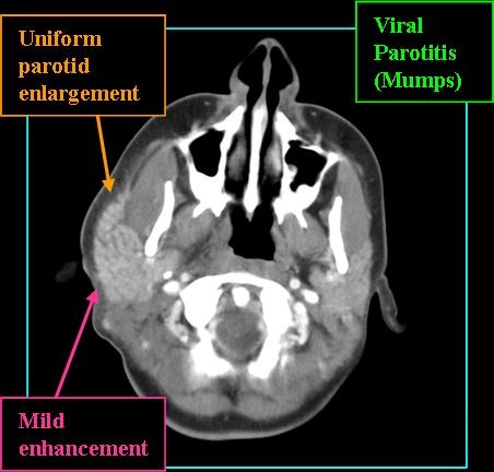
- Viral - acute viral parotitis with systemic infection; most commonly mumps paramyxovirus, sometimes influenza and Coxsackie virus
- Calculus-induced - parotiditis secondary to duct obstruction from sialolith
- Commonly unilateral with overlying soft tissue involvement
- Viral parotitis is often bilateral, however one gland is affected 1-5 days before the contralateral gland
Radiologic Findings
- Look for radiopaque calculus with duct dilatation on non-contrast CT with calculus-induced parotitis
- Gland should be diffusely and uniformly enlarged
- Contrast-enhanced CT of bacterial parotitis will show enhancing gland with subcutaeous fat-stranding
- MR signal high on T2 and post-contrast T1 shows gland enhancement
- Look for ring-enhancing lesions and soft tissue edema suggestive of abscess formation (Patel 2023)
- Sialography is contraindicated in acute infection due to risk of contrast extravasation and potential to cause pain or damage to glandular tissue (Patel 2023)

References
Patel J, Maymeskul V, Kim J. Infections of the Oral Cavity and Suprahyoid Neck. Oral Maxillofac Surg Clin North Am. 2023 Aug;35(3):283-296. doi: 10.1016/j.coms.2023.01.001. Epub 2023 Apr 7. PMID: 37032180.
Pollenus J, Van Lierde S. Neonatal Parotitis: A Case Report and Review of the Literature. Pediatr Infect Dis J. 2023 Sep 1;42(9):e323-e327. doi: 10.1097/INF.0000000000003959. Epub 2023 May 5. PMID: 37171966.
Henry T Hoffman, MD, MS, FACS SECTION EDITOR: Daniel G Deschler, MD, FACS DEPUTY EDITOR: Jane Givens, MD, MSCE. Salivary Gland Swelling Evaluation and Management. UpToDate March 2024.