

 and dilation with rigid Jackson laryngeal dilator")


Click on photos above to enlarge
Return to: Subglottic Stenosis Protocol
other examples: Case Example 1: Subglottic Stenosis due to Wegener's Granulomatosis
Case Example Subglottic Stenosis and Relapsing Polychondritis
Case Example Posterior Scar Band
Idiopathic (? reflux related) Subglottic Stenosis Example Case
Case #1 57 yo female with progressive shortness of breath beginning in 2001 with treatment for 'idiopathic subglottic stenosis' employing flexible fiberoptic Yag laser radial cuts and balloon dilation iniitally Nov 2001 (by pulmonary medicine under conscious sedation) with three subsequent treatments, the most recent being November 2008 (Andrews BT et al). She was re-evaluated in Otolaryngology on Sept 9 2009 with progressive exertional dyspnea.
- A remote history of previous esophagoscopy 'showing reflux' with continued treatment with omeprazole 20 mg po qd
- Evaluation by Oto on Sept 2009: negative autoimmune w/u with transnasal laryngoscopy:
Operative intervention 10-21-09 (mask anesthesia with full relaxation progressing to jet anesthesia followed by intubation):
Placement of the Dedo Laryngoscope positioned with the tip slightly separating the posterior vocal fold to permit photography, insufflation of 1 cc of 4% lidocaine and then application of jet anesthesia.
Initiatiating treatment: Injection of the subglottic narrowing is done with a kenalog 10 preparation made by diluting kenalog 40 (1 part) with 1% lidocaine, 1:100,000 epinephrine (3 parts). Scissors incision on either side of a 3 mm segment of the stenosis at 12:00 (anterior) permits a biopsy to be taken. Radial cuts are generally made then at 3:00 (right lateral) and 9:00 (left lateral) with scissors.
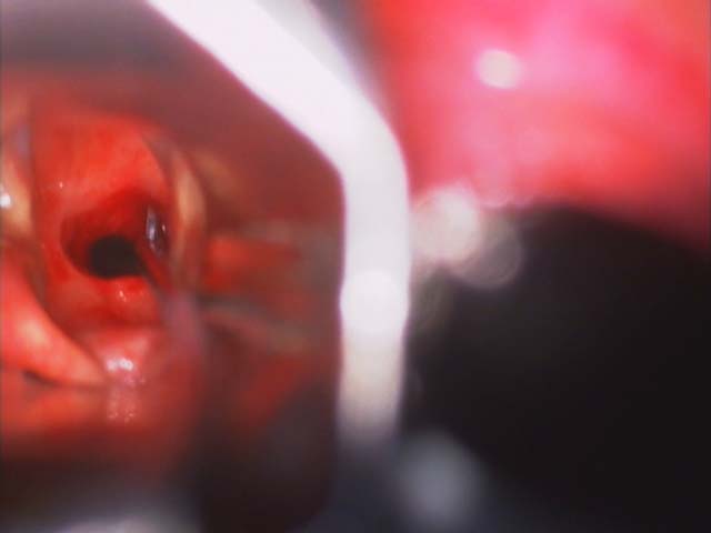
Initial exposure:
Injection with 'kenalog 10' see dilution above:
Scissors incision: after biopsy (acute inflamation and fibrosis) and dilation with rigid Jackson laryngeal dilator:
Room set up with endoscopy during balloon dilation:

Measurement of the stenosis: The location of the cuts may be modified by the nature of the narrowing. Extent of narrowing is measured by placing a laryngeal suction with its tip at the level of the vocal cords. A mark is made on the suction with a pen where it abuts the entrance to the Dedo laryngoscope. The suction tip is then advanced to the near (superior) level of the stenosis prompting placement of a second pen mark on the suction where it now abuts the entrance to the Dedo laryngoscope. The suction tip is then advanced to the far (inferior) end of the stenosis prompting placement of the a third mark. The distance between the marks on the suction is then measured and recorded - between the 1st and 2nd mark = distance stenosis begins below the free edge of the vocal cord
distance between 2nd and 3rd mark = length of stenosis
Video of dilation - metal Jackson laryngeal dilator:
Initial dilation is done with the metal Jackson laryngeal dilators. This part of the procedure may precede the injection, biopsy, radial cuts and measurements above if the narrowing is sufficient to compromise the airway. These dilators are used primarily (in the small female larynx) to improve the airway sufficiently to permit placement of a small endotracheal tube (4-0 MLT or 5-0 MLT) to permit balloon dilation. The risk of injury to the small female larynx by passing rigid metal dilators larger than 36 is diminished by this approach. The large male larynx will more readily accomadate the larger 40 to 42 metal Jackson dilators. It is difficult to safely place a dilator larger than 42 through a Dedo laryngoscopy (sometimes may need to stop at 40).
Subglottic Stenosis/SGS dilator 102109
Video of dilation - CRE 15 - 18 mm balloon dilation
With a small endotracheal tube in place, dilation with the CRE balloon dilating system may proceed safely with continued ventilation and diminished risk of injury to the glottic larynx:
(see also: Subglottic Stenosis - Upper Tracheal Stenosis CRE Balloon Dilation)
Subglottic Stenosis/SGS balloon dilation 102109
Esophagoscopy (GE junction): acute inflamation
References
Hulstein S, Hoffman H. Technique for improved safety in the endoscopic management of subglottic stenosis. Am J Otolaryngol. 2016 Nov - Dec;37(6):490-492. doi: 10.1016/j.amjoto.2015.10.009. Epub 2015 Nov 10.
Andrews BT, Graham SM, Ross AF, Barnhart WH, Ferguson JS, and McLennan G: Technique, utility, and safety of awake tracheoplasty using combined laser and balloon dilation. Laryngoscope. 2007 Dec;117(12):2159-62
Shapshay SM, Bellapianta KM: letter to the editor in reference to "Andrews BT, Graham SM, Ross AF, Barnhart WH, Ferguson JS, and McLennan G: Technique, utility, and safety of awake tracheoplasty using combined laser and balloon dilation. Laryngoscope. 2007 Dec;117(12):2159-62" Laryngoscope 2008 Jun;118(6):1133-4
Informed consent approving "use of photos, videos, case history for presentation, publications, purposes in any media" on file.