See also:
- Anatomy of submandibular gland and duct
- Plunging Ranula Transoral Resection (Sublingual Gland) Aided With Sialendoscopy with Histopathology
Uncinate Process of the Deep Lobe of the Submandibular gland
The normal shape of the submandibular gland is broadly considered ellipsoid despite the description in the 1985 version of Gray’s Anatomy as ‘round and about the size of a walnut’ (Gray H 1985). This text describes the gland in context of superficial and deep surfaces but isolates only a single subunit termed a ‘deep process’ of the gland extending anteriorly between the mylohyoid muscle laterally and the hyoglossus and styloglossus muscles medially. Other anatomists identify this extension as a ‘deep portion of the gland …typically folded around the posterior edge of the [mylohyoid] muscle.’ (Hollinshead 1968). This deep portion of the gland where it ‘wraps around the posterior edge of the mylohyoid muscle’ has also been termed ‘the uncinate process’ (Xu and Chang 2021).
Sabin et al divided the gland into ‘two sets of lobes: superficial and deep” (Saban 2021). These investigators identified the paired submandibular glands as being like “elongated, flattened hooks” with the deep lobe hooking around the posterior margin of the mylohyoid muscle. They compared the submandibular gland to a “triangular almond” whose anterior border is divided by the mylohyoid muscle to define the two lobes – superficial and deep. Although the mylohyoid is used by multiple investigators to distinguish the superficial lobe (above the mylohyoid) from the deep lobe (superficial, under the skin), these assessments fail to define the extent of separation into lobes posterior to the free edge of the mylohyoid (Atkinson 2018, Miloro 2019).
Uncinate (Merriam-Webster 2023) "bent at the tip like a hook" etymology: (Latin) 'uncinatus from uncinus hook, from uncus'. The uncinate process within the sino-nasal region is part of the osteomeatal complex with variable anatomy as a 'thin, semi-circular bony process of variable length' located 'either frontally and inferior, or anteriorly and supreiorly to the inferior turbinate's ethmoidal process" (Güngör 2016). The uncinate processes within the cervical spine are projections at the level of the third cervical vetebra that may extend as low as the second thoracic vertebra (Hartman 2014). The uncinate process of the pancreas is one of 4 segments - defined as 'the extension of the medial-caudal portion of the head" with a 'concave anteromedial border and triangular pattern"(Dilek 2021).
Ultrasound of the Submandibular Gland
Case Example
34 year old female with history of swelling in right submandibular area for 1 1/2 years without fluctuation and no dry mouth was evaluated with CT imaging identifying ranula
Histopathology
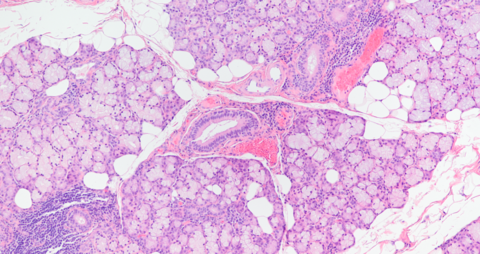
Submandibular gland histologically showing serous-dominant acini consistent with submandibular gland (evaluated in full with no region of mucus-dominant acini as from sublingual gland)
Resection permitted histopathologic analysis with the resected sublingual gland showing abundant mucinous cells in the specimen as expected (sublingual gland predominantly mucinous with serous demilunes at the periphery of scattered mucinous acinar cells)


Submandibular Gland 12.5x

Submandibular Gland 40x

Submandibular Gland 100x

References
Katz P, Hartl DM, Guerre A. Clinical ultrasound of the salivary glands. Oto Clin N Am. 2009; 42(6):973-1000
Koch M, Mantsopoulos K, Leibl V, Müller S, Iro H, Sievert M. Ultrasound in the diagnosis and differential diagnosis of enoral and plunging ranula: a detailed and comparative analysis. J Ultrasound. 2022 Dec 17. doi: 10.1007/s40477-022-00743-7. Epub ahead of print. PMID: 36527568.
Jain P, Jain R. Types of sublingual gland herniation observed during sonography of plunging ranulas. J Ultrasound Med. 2014 Aug;33(8):1491-7. doi: 10.7863/ultra.33.8.1491. PMID: 25063415.
Gray, Henry. Anatomy of the Human Body 30th American Ediition edits by Clemente CD Lea & Febiger 1985 PhiladelphiaCh 16 The Digestive System Submandibular Gland p 1434
Hollinshead WH: Anatomy For Surgeons: Volume 1 The Head and Neck 2nd edition Harper & Row Publishers Cambridge 1968pp 428-9 Chapter 7: The Jaws, Palate, and Tongue
Xu MJ and Chang JL: Practical Salivary Ultrasound Imaging Tips and Pearls. Otolaryngol Clin N Am 54 (2021) 471-487 Elsevier
Saban Y, Tevfik S, Palhazi P and Polselli R: “Salivary Gland Anatomy” Chapter 1 pp 2-11 in Surgery of the Salivary Glands ed Robert Witt 2021 Elsevier Inc. Edinburgh
Atkinson C, Fuller J 3rd, Huang B. Cross-Sectional Imaging Techniques and Normal Anatomy of the Salivary Glands. Neuroimaging Clin N Am. 2018 May;28(2):137-158. doi: 10.1016/j.nic.2018.01.001. Epub 2018 Mar 7. PMID: 29622110.
Miloro M and Kolokythas A: Diagnosis and Management of Salivary gland Disorders – Chapter 21, pp 423-449 in Contemporary Oral and Maxillofacial Surgery Deditor James R Hupp MD JD MBA 2019 Elsevier, Inc
Jain P. High resolution sonography of sublingual space. J Med Imaging Radiat Oncol 2008; 52:101–108
Jain P, Jain R, Morton RP, Ahmad Z. Plunging ranulas: high-resolution ultrasound for diagnosis and surgical management. Eur Radiol 2010; 20:1442–1449.
Merriam-Webster.com Dictionary, Merriam-Webster,“Uncinate.” https://www.merriam-webster.com/dictionary/uncinate. Accessed 3 Apr. 2023.
Güngör G, Okur N, Okur E. Uncinate Process Variations and Their Relationship with Ostiomeatal Complex: A Pictorial Essay of Multidedector Computed Tomography (MDCT) Findings. Pol J Radiol. 2016 Apr 20;81:173-80. doi: 10.12659/PJR.895885. PMID: 27158282; PMCID: PMC4841358.
Hartman J. Anatomy and clinical significance of the uncinate process and uncovertebral joint: A comprehensive review. Clin Anat. 2014 Apr;27(3):431-40. doi: 10.1002/ca.22317. Epub 2014 Jan 22. PMID: 24453021.
Machado MA, Ardengh JC, Makdissi FF, Machado MC. Minimally Invasive Resection of the Uncinate Process of the Pancreas: Anatomical Considerations and Surgical Technique. Surg Innov. 2022 Oct;29(5):600-607. doi: 10.1177/15533506211045317. Epub 2022 Mar 25. PMID: 35332821.
Dilek O, Akkaya H, Kaya O, Inan I, Soker G, Gulek B. Evaluation of the contour of the pancreas: types and frequencies. Abdom Radiol (NY). 2021 Oct;46(10):4736-4743. doi: 10.1007/s00261-021-03152-2. Epub 2021 May 31. PMID: 34057566.
Kiesler K, Gugatschka M, Friedrich G. Incidence and clinical relevance of herniation of the mylohyoid muscle with penetration of the sublingual gland. Eur Arch Otorhinolaryngol. 2007 Sep;264(9):1071-4. doi: 10.1007/s00405-007-0321-1. Epub 2007 May 4. PMID: 17479273.
Ab Rahim NAC, Liew YT, Ghauth S, Narayanan P, Abu Bakar Z. A Single Institution Cadaveric Study on Anatomical Variation of the Sublingual Gland Duct. Indian J Otolaryngol Head Neck Surg. 2023 Jun;75(2):347-351. doi: 10.1007/s12070-022-03261-4. Epub 2022 Nov 7. PMID: 36406798; PMCID: PMC9640814.8


