Carver College of Medicine to purchase Cryo-EM instrument
Plans are underway to enhance structural biology capabilities at the University of Iowa Roy J. and Lucille A. Carver College of Medicine by purchasing a cryo-electron microscope. The new instrument, a 200 kV Glacios Cryo-TEM microscope from ThermoFisher Scientific, will facilitate the study of large and complex biological structures, such as proteins, viruses, and cellular components, which are difficult or impossible to visualize by other methods.
The Glacios microscope is anticipated to arrive in fall 2025 and will be housed in the Protein and Crystallography Facility.


Better images are revolutionizing structural biology
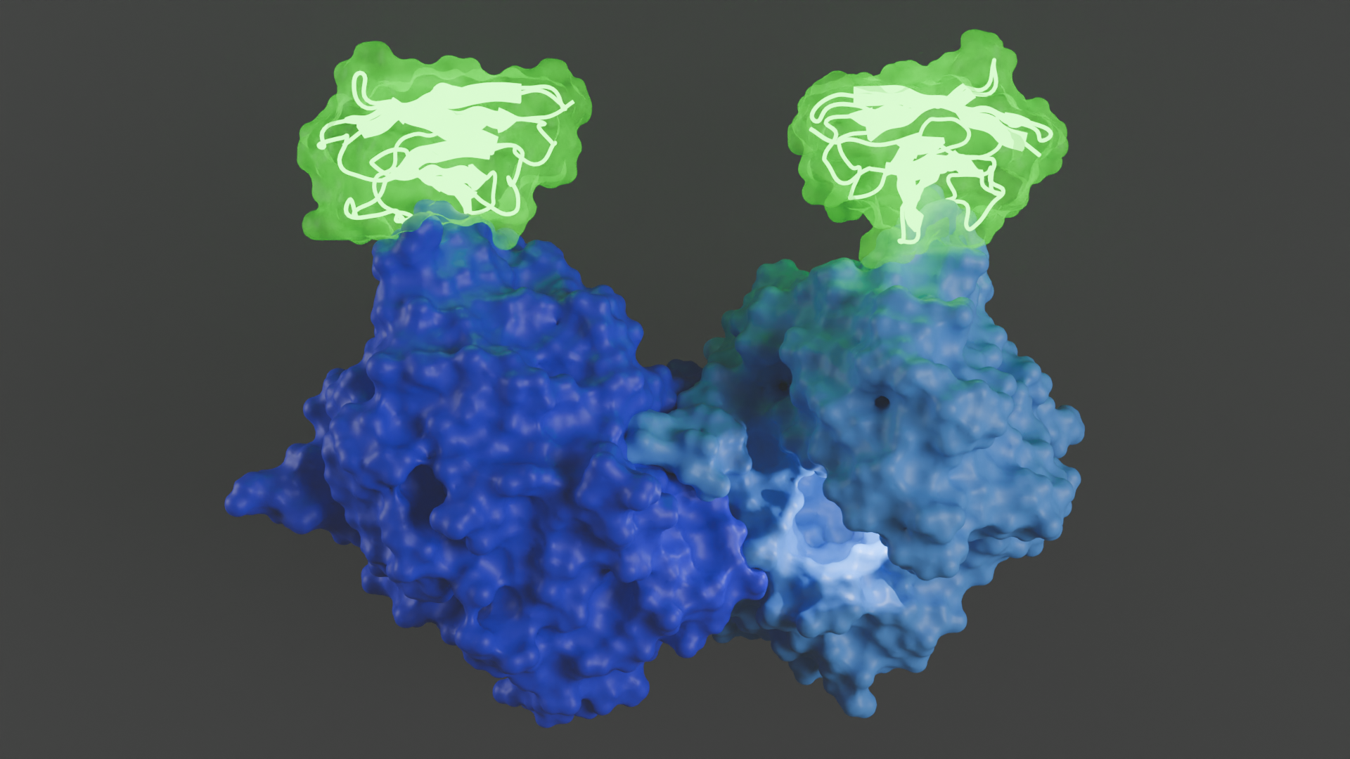
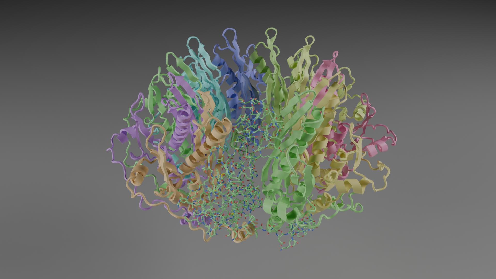
Cryo-electron microscopy (Cryo-EM) is an advanced imaging technique used to study the 3D structures of biological molecules in a near native environment at the atomic level. In cryo-EM, samples are rapidly frozen in vitreous ice to preserve their natural state and prevent damage from the electron beam. The technique produces higher resolution images and requires much less sample than other structural methods like X-ray crystallography and NMR. The images are then combined and processed using computational methods to create a detailed 3D model of the molecule or complex being studied.
Nick Schnicker, PhD, director of the UI Protein and Crystallography Facility, notes that the structures produced by cryo-EM provide detailed insights into how biological molecules function, how they interact, and how they might be targeted for drug development, contributing to advancements in medicine and understanding of biological processes.
“Cryo-EM has revolutionized the field of structural biology and has become an essential tool for scientific research in numerous disciplines,” he says. “Acquiring the Glacios microscope will transform our cryo-EM capabilities at the University of Iowa and keep our researchers at the forefront of scientific research and innovation.”
UI researchers currently have access to cryo-EM microscopy through the Protein and Crystallography Facility’s network with both regional and National Centers for Cryo-EM but having the technology in-house will increase access and training opportunities according to Schnicker.
“In addition to advancing biomedical research and promoting collaboration between researchers and across disciplines, having direct access to cryo-EM instrumentation will help attract talented, innovative researchers and students looking to tackle challenging scientific questions,” he says.
The purchase of the Cryo-EM instrument is one example of how the UI Carver College Medicine invests in research resources to provide a strong collaborative research environment that enables high quality science.
“Providing access to the most advanced scientific technology and instrumentation is critical for a thriving research ecosystem,” says Robert Piper, PhD, associate dean for research, and director of the Core Research Facilities at the Carver College of Medicine. “Equipping our scientists and trainees with the advanced tools and hands-on experience they need, allows them to forge new research ventures, make exciting discoveries, and remain competitive for extramural funding.”

| Electron source | 200kV, XFEG |
| Sample exchanger | 12 grid autoloader |
| Stage | ±70° tilt |
| Software | Smart EPU w/QM, EPU multigrid |
| Computing | Data management platform |
| Optics | Fringe-free imaging |
| Cameras | Falcon 4i, Ceta-D |
| Energy filter | Selectris |
| Resolution | < 3 A |