A new gallery of biomedical research images is now on display on the ground floor of the Carver Biomedical Research Building. The images, each measuring 5 feet in diameter and mounted on back-lit frames, are visible to pedestrian and vehicle traffic along Newton Road. The gallery highlights the breadth of foundational research conducted in the Carver College of Medicine.
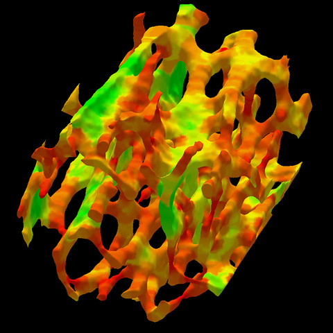 Identifying osteoporosis

Osteoporosis is a common and debilitating condition of aging. Treatments are most effective in the early stages of the disease. Therefore, identifying early bone loss is a key to slowing and/or stopping disease progression. Bone loss begins in the trabecular or “spongy” bone, which is found at the ends of the cortical bones that provide structural support for the body. This image shows the plates (green) and rods (red) of human trabecular bone at the distal tibia. Histologic evidence confirms the relationship between the gradual conversion of trabecular plates to rods and increased fracture risk. The Saha Lab developed a computational method that identifies these structures more distinctly than other imaging options. The unique algorithm characterizes individual trabecular bone on the continuum between a perfect plate (green) and perfect rod (red), enabling early detection of imbalance between trabecular bone plates and rods and thus, increased risk for fracture.
Punam Saha, PhD
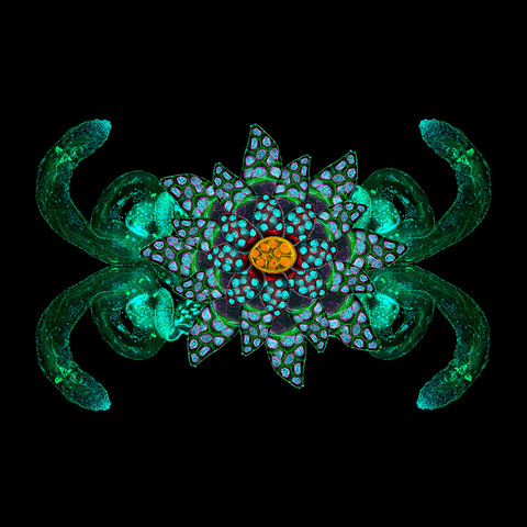 Biology of cancer

In astrology, the zodiac sign for cancer is a crab. This crab-like shape is a Drosophila, or fruit fly, reproductive system presented in mirror reflection. The Tootle Lab is using the Drosophila ovary to to better understand the fundamental biology of cancer. They are investigating the interplay between prostaglandins and lipid droplets in remodeling the actin cytoskeleton. Misregulation of prostaglandins and lipid droplets have both been implicated in human cancer. Likewise, remodeling of the actin cytoskeleton is required for reproduction, development and disease, and both are known to have independent roles in cancer. This research project is exploring a novel pathway involving a previously unknown connection between prostaglandins and lipid droplets in the regulation of actin dynamics. Changes in regulation of actin dynamics are implicated in some cancers. This work may reveal new therapeutic targets as scientists begin to understand the relationship between prostaglandins, lipid droplets, and cytoskeletal remodeling.
Michelle Giedt, PhD
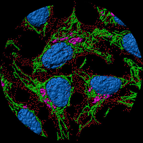 Packing, sorting and delivering insulin in cells

Diabetes interferes with the ability of pancreatic beta-cells to package and release insulin. Understanding how insulin is produced, packaged, stored and released is the primary focus in the Stephens lab. Each tiny red dot in this image represents a package of insulin ready to be sent out of the pancreas. Using microscopy to follow these packages from development to delivery allows direct observation of the differences in function between healthy and diabetic cells. Additional structures in this image include mitochondria (green), Golgi (magenta), and nuclei (blue). Dr. Stephens is a member of the UI Fraternal Order of Eagles Diabetes Research Center, where dozens of scientists are focused on identifying the biological underpinnings of the disease and developing new therapeutics.
Sam Stephens, PhD
Fraternal Order of Eagles Diabetes Research Center
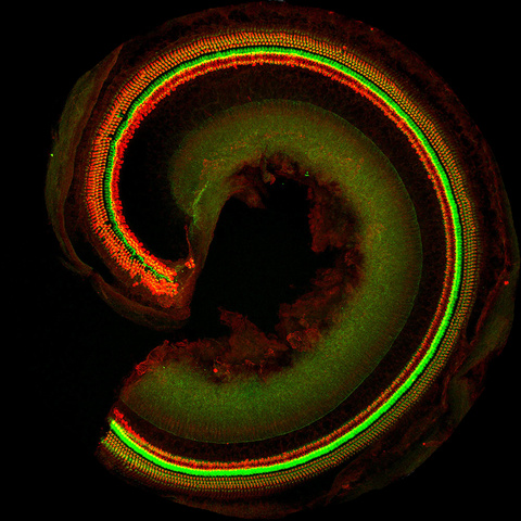 Gene therapy for hearing loss

The cochlea is a key part of the inner ear that converts waves generated by sounds into electrical signals that travel to the brain. This immunofluorescence of a mouse cochlea was captured by confocal microscopy after dissecting the membranous labyrinth from the temporal bone. The bright red curved structure shows part of a spiral shaped cochlea after it was removed from the temporal bone. The green stripe that separates the inner hair cells from the outer hair cells highlights the filamentous actin in the hair cells and pillar cells. One goal of scientists in the Molecular Otolaryngology & Renal Research Laboratories (MORL) is to develop gene therapy for hearing loss by transducing hair cells with vectors carrying normal genes that can be used to replace defective genes. Cochlear immunohistochemistry allows scientists to quantitate transduction efficiency as one method for evaluating therapeutic effect.
Hidekane Yoshimura, PhD
Molecular Otolaryngology and Renal Research Laboratories
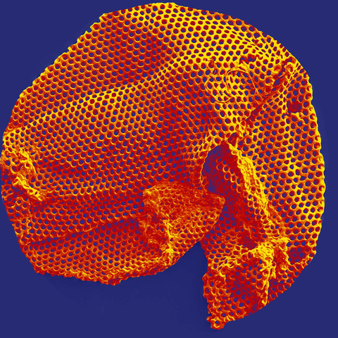 Supporting vision restoration

This scaffold may hold the key to restoring lost vision for some patients. It is used to support the attachment and growth of retinal progenitor cells (stem cells) during retina transplant. To date, it has shown success in small- and large-animal models of retinal degeneration. UI biomedical engineers are working to optimize these scaffolds – design/structure, degradation rate, thickness, and stiffness – for human transplant. They have developed a simple, rapid photopolymerization technique that produces thin, porous, degradable films, like the one pictured here. The material is so delicate that it folds in on itself during preparation for the scanning electron microscope (SEM) imaging.
Kristan Worthington, PhD
Institute for Vision Research
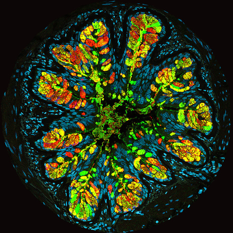 Understanding mucus

Airways secrete mucus to protect the lung from environmental injury caused by pathogens and particles in inhaled air. However, in diseases such as asthma, cystic fibrosis, and chronic obstructive pulmonary disease (COPD), mucus hypersecretion and accumulation obstruct the airway and limit breathing. The image shows mucus hypersecretion and accumulation in an asthmatic distal airway. Airway cells are labeled in light blue with nuclear staining. Goblet cells in airway produce two types of mucin proteins, MUC5AC (red) and MUC5B (green); they are the most important components of airway mucus. The airway lumen is collapsed and filled with mucus instead of air. Scientists and physicians in the Lung Biology and Cystic Fibrosis Research Center are working to understand the biochemical and biophysical properties of mucus and to develop novel treatments for people who have cystic fibrosis and other airway diseases.
Wenjie Yu, PhD
Lung Biology & Cystic Fibrosis Research Center
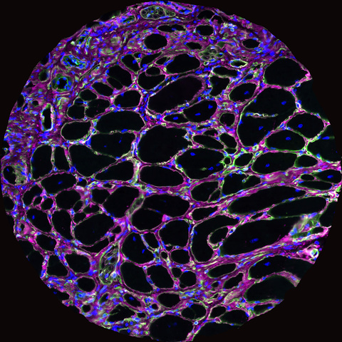 Muscle fibers and fibrous tissue in muscular dystrophy

This is an image of mouse muscle that has undergone immunofluorescent stain, which produces the bright colors. Immunofluorescent staining helps scientists determine whether particular proteins are present in the tissue. In this image, proteins are stained to highlight muscle fibers and fibrous tissue, which allows researchers to measure the diameter of the fibers and amount of fibrous tissue. These are indicators of muscle health--small fibers and high fibrous content indicates weakened muscle. The Campbell Lab has been working for more than two decades to identify the mechanisms that cause muscular dystrophy and to develop therapies for these diseases.
Megan Devereaux
Wellstone Muscular Dystrophy Cooperative Research Center
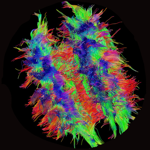 Wiring of the human brain

The intricate connections between nerve cells in the brain form the basis for our thoughts, behaviors and actions. Knowledge of the complex networks in the brain is crucial to understanding the structural and functional organization of the brain. The image is a visualization of the wiring of the human brain. Research in the Magnotta Lab focuses on developing novel imaging techniques that can elucidate more details of this wiring such as its complex crossings and turnings. Widespread or spatially localized degeneration of the wiring in response to an underlying pathology or aging can lead to loss of brain function. Researchers anticipate that images like these will lead to development of spatially targeted regenerative interventions to restore the wiring and restore brain function in such cases. This image was collected on the Iowa Magnetic Resonance Research Core Facility’s 7T MRI scanner.
Merry Mani, PhD
Iowa Neuroscience Institute
Iowa Institute for Biomedical Imaging