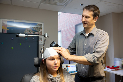
Deep brain electrodes measure effects of transcranial magnetic stimulation in real time
Researchers at the University of Iowa and Stanford University have developed a new tool that allows scientists to safely and accurately measure the effect of transcranial magnetic stimulation (TMS) on the activity of deep brain structures. The new technique, known as TMS-iEEG (intracranial electrocorticography), is providing hard data on how TMS works and may lead to improvements in the technology that is currently used to treat several neuropsychiatric conditions.
TMS, which delivers magnetic pulses non-invasively to the brain from the surface of the head, is approved to treat depression, obsessive-compulsive disorder (OCD), migraine, and smoking cessation, but doctors do not have a clear idea of how it works.
“People assume that TMS works by modulating the region of the cortex that you're targeting, and then that subsequently modulates the deeper regions that are functionally connected to the original targeted area,” explains Aaron Boes, MD, PhD, UI associate professor of pediatrics, neurology, and psychiatry. “That has been an assumption without a lot of great data to back it up.”
The new TMS-iEEG technique developed by Boes and his colleagues at UI and Stanford is now providing that data and revealing a clearer picture of how the brain responds to TMS in real time—by recording directly from within the brain of neurosurgery patients implanted with electrodes while they receive TMS. Overall, the findings, which were published recently in Molecular Psychiatry, suggest that TMS-iEEG is a viable tool for studying the electrophysiological effects of TMS on the human brain in real time, including which deep brain structures are actually being modulated by clinical TMS protocols.
UI’s collaborative environment yields results


Boes first became interested in understanding how TMS exerts its clinical effect while he was at Harvard. Prompted by discussions with his mentor Mike Fox, he became fascinated by the idea of measuring how TMS stimulation of the cerebral cortex provokes activity in deeper brain structures through network connectivity pathways.
Moving to Iowa in 2017 provided Boes with the opportunity to pursue the idea. He teamed up with Matthew Howard, MD, PhD, professor and chair of neurosurgery, whose team is expert in using deep brain electrodes to capture neural activity from patients undergoing brain mapping for epilepsy surgery. The UI researchers, along with Corey Keller’s team at Stanford, set about combining their unique technologies to study the effects of TMS in real time.
First came the crucial, painstaking safety testing to ensure that combining TMS with implanted electrodes in a human brain would not cause harm. Led by Hiroyuki Oya, MD, PhD, UI professor of neurosurgery, the researchers used a gel-form replica of a brain to prove that the TMS magnetic field did not cause implanted electrodes to move, heat up, or generate a current at the electrode site.
Once the technique had passed these safety tests, the researchers conducted human studies in a small group of patients with electrodes implanted to pinpoint seizure-inducing brain sites for removal. Over the next several years, the team accrued sufficient data to show that TMS is capable of inducing responses both in the areas of the cortex that are being directly stimulated and also in functionally connected deep brain structures in humans. For example, the team showed a strong and consistent activation in the dorsal anterior cingulate, a region located deep in the middle of the brain; an effect that would not have been measurable using traditional external EEG.
“It is really hard to record from these deep brain structures on a time scale that would be reasonable to look at the effects of TMS, which is less than a second,” Boes says. “Functional MRI, which is often used to measure brain connectivity, is too slow to capture the effect of TMS. In contrast, intracranial electrocorticography (iEEG) is fast enough to measure brain activation at the deep structures on the same time scale as TMS works.”
The technique can also measure how TMS changes the brain by measuring excitability before and after TMS treatment at specific regions.
New frontiers for TMS
Boes and Keller recently received funding from the National Institutes of Health to extend their findings by using TMS-iEEG to measure the effects of a new TMS protocol for depression treatment. The new protocol uses repeated TMS sessions that essentially deliver a traditional six-to-eight-week treatment course of TMS in a single day, and then repeating it for five consecutive days.
In addition to Boes’ research focus on the effects of the new TMS approach, patients at UI will also have access to this new TMS treatment through a new clinical trial headed by Nick Trapp, MD, UI assistant professor of psychiatry.
“It is something we’re excited about in the TMS world based on some promising clinical trial results,” says Boes, who also is director of the Iowa Center for Noninvasive Brain Stimulation and a member of the Iowa Neuroscience Institute. “Both the clinical trial and our continued research efforts are about pushing the envelope to better understand how TMS works in order to improve upon existing TMS treatments for depression and other neuropsychiatric conditions.”
The study was funded in part by grants from the National Institute of Mental Health, and the National Institute of Neurological Disorders and Stroke, and the Roy J. Carver Charitable Trust. UI researchers Joel Bruss and Phillip Gander were also part of the team.