UI researchers led by Sam Young, PhD, have just published findings from a study using innovative technologies to better understand signaling capacity between neurons, leading to a new understanding of how the strength of information flow in the brain is controlled.
First, some definitions
The fundamental process of information transfer from neuron to neuron occurs through a relay of electrical and chemical signaling at the synapse, the junction between neurons. Electrical signals, called action potentials, cause voltage-gated calcium channels on the presynaptic neuron to open. The influx of calcium through the channels triggers the release of neurotransmitters (the chemical messengers), which travel across the synapse to the next neuron in the relay, passing along the information.
“The mechanisms involved in controlling the strength of the information flow are critically important for brain function,” says Young, associate professor of anatomy and cell biology at the UI Carver College of Medicine and a member of the Iowa Neuroscience Institute.
Study asks: Can new channels be added?

“Critical to controlling information flow is the influx of calcium through calcium channels at the synapses, and that depends on the number of calcium channels embedded in the presynaptic membrane. Despite their importance, we don’t understand what controls the number of calcium channels at the synapse,” Young says.
In the central nervous system, there are three subtypes of the Cav2 family of voltage-gated calcium channels. Of these, Cav2.1 is the most efficient at triggering neurotransmitter release from the synapse since it is present at higher levels compared to other Cav2 subtypes. The Cav2.1 subtype also is the dominant isoform associated with human diseases known as Cav2 channelopathies, which include forms of migraine, epilepsy, and ataxia.
A better understanding of how Cav2.1 and other calcium channels involved in neurotransmission are regulated might reveal ways to therapeutically strengthen synapses that are failing in a disease state.
Questioning a current assumption
Current dogma in the field has proposed that the active zone, the specialized site on the synapse that controls neurotransmitter release, can only accommodate a specific number of Cav2 calcium channels and is always filled to capacity. In particular, this suggests it is not possible to increase the number of Cav2.1 channels. Therefore this limits the means by which synaptic strength can be increased.
“We were able to show that actually you can put in more Cav2.1 channels in the active zone indicating the active zones are not filled to capacity. Moreover, adding more channels increases synaptic strength,” says Young, who was senior author of the study, which was published Dec. 10 online in the journal Neuron.
The team also showed that these findings were true for their neuronal circuit at both early and mature developmental time points.
“That means that the ability to put in more calcium channels is ongoing through the lifetime of the organism ,” Young adds.
Innovation drives discovery
Young and his team made their discoveries using several innovative technologies that they have developed.
For example, a gene therapy vector called helper dependent adenoviral vector, which has a larger carrying capacity than more traditional vectors, allowed them to deliver the relatively large genes encoding the calcium channels into a native intact neuronal circuit.
They also have a system that allows for overexpression of the calcium channels in neurons. Together these two technologies allowed the team to force overexpression of the different calcium channel subtypes to investigate the effects on synaptic strength.
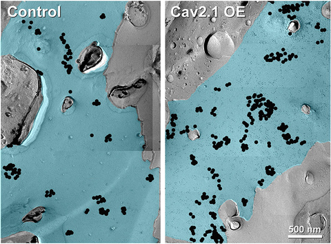
In addition, the team used an imaging technique that labels individual calcium channels with gold and allows the scientists to visualize the calcium channels and physically quantify the number of channels in the active zone.
“This technique allowed us to show that the changes were really due to an increase in the number of channels because we could directly visualize the individual calcium channels and measure them,” Young says.
Study finding: Number of calcium channels can increase

The image shows gold-labelled Cav2.1 calcium channels (black dots) in the plasma membrane of the Calyx of Held nerve terminal. The image on the left is the control. The image on the right shows that overexpression of the Cav2.1 alpha 1 subunit leads to an increased number of Cav2.1 channels, indicating that the active zone is not filled to capacity and that it can accommodate additional Cav2.1 channels.
The findings suggests it is possible to increase the number of calcium channels in the active zone and that this is a way to strengthen neurotransmission between neurons.
“Now we know there must be specific mechanisms either at the synapse or at some other checkpoint that are controlling the ratio and type of channels that are used. But what those mechanisms are is a big unsolved question,” Young says. “Basically, this study opens up a whole new avenue of research and new ways of thinking about how synapses regulate synaptic strength. Furthermore, these findings will provide fundamental insights into how information is encoded by the nervous system, facilitating the development of treatments for a wide range of neurological and neuropsychiatric disorders.”
Members of the research team
In addition to Young, the research team included R. Oliver Goral and Christian Keine in the UI Department of Anatomy and Cell Biology; former members of Young’s laboratory at the Max Planck Florida Institute for Neuroscience: Matthias Lubbert, Travis Putzke, and Rachel Satterfield: and members of Max Planck Florida Institute for Neuroscience Electron Microscopy Core, Connon Thomas, Debbie Guerrero-Given, and Naomi Kamasawa.
This work was supported by a research grant from the National Institute of Deafness and Communication Disorders, part of the National Institutes of Health (R01 DC014093), the University of Iowa, and the Max Planck Society.