

Click on image to enlarge, advance with cursor over right lateral border
see also: Instructions to patients submandibular salivary stones; Case Example 2 Retrograde Sialendoscopy to Prevent Retained Ductal Stone with Submandibular Gland Resection
return to: Submandibular Gland Resection or Sialolithiasis
Case Example
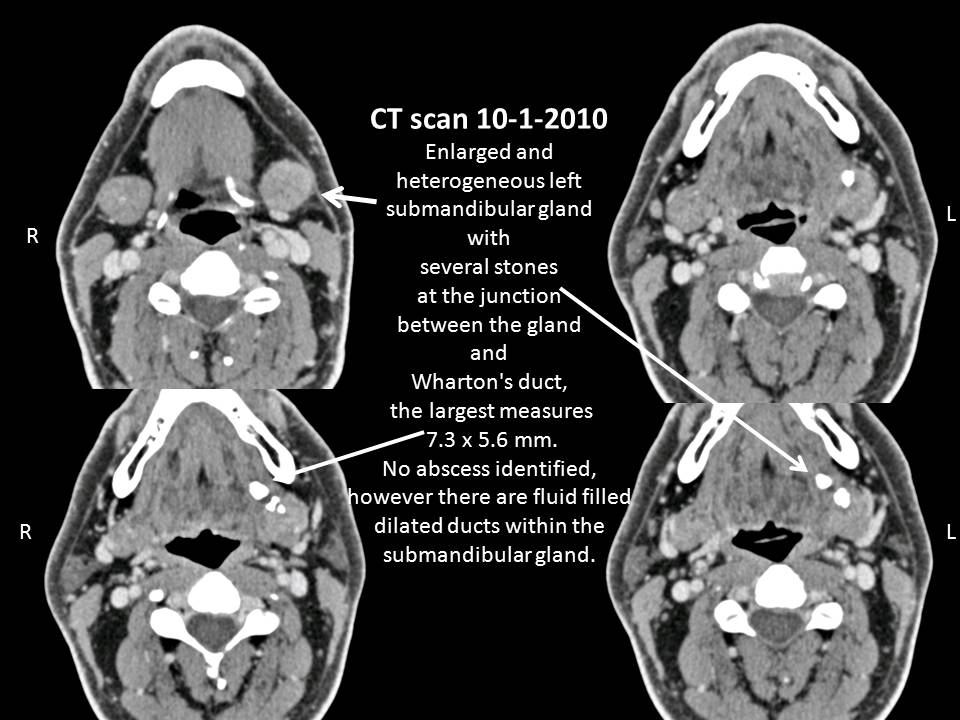
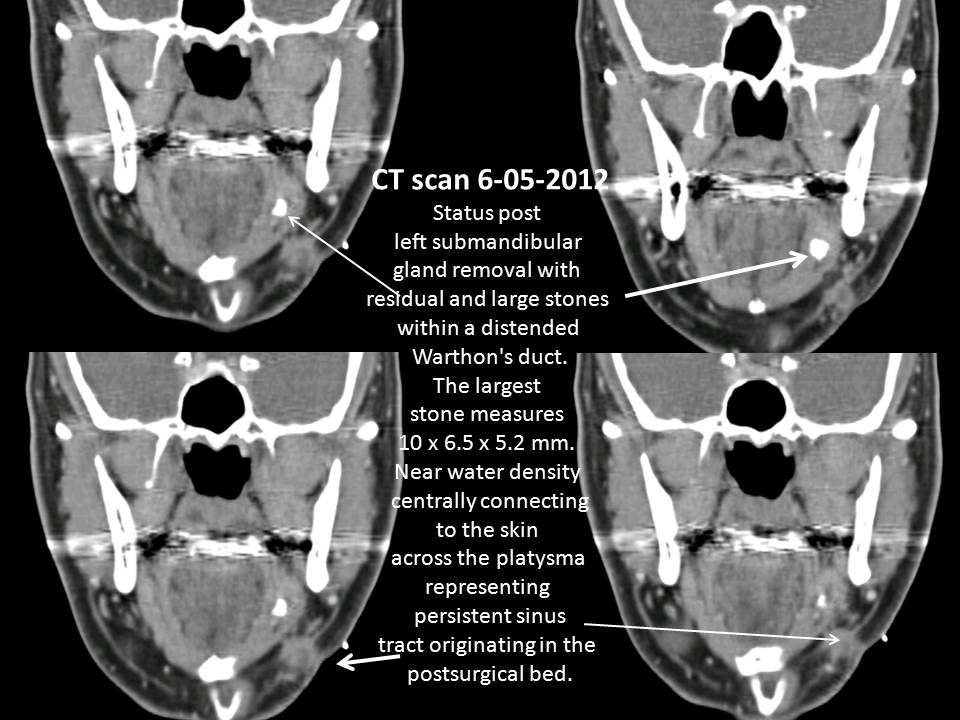
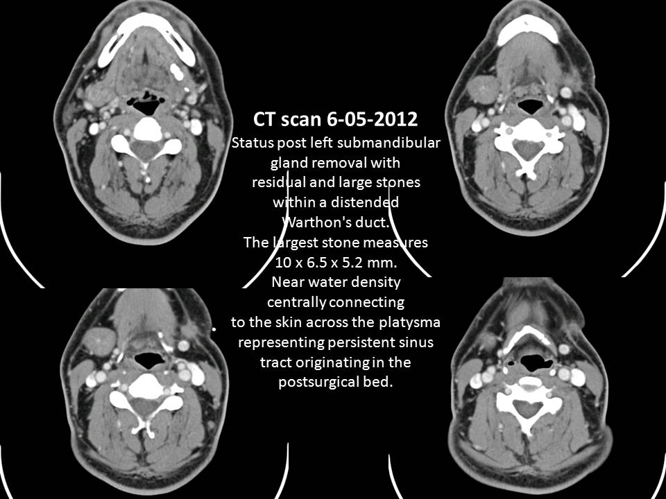
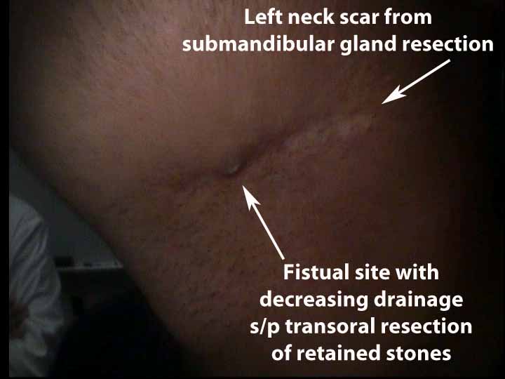
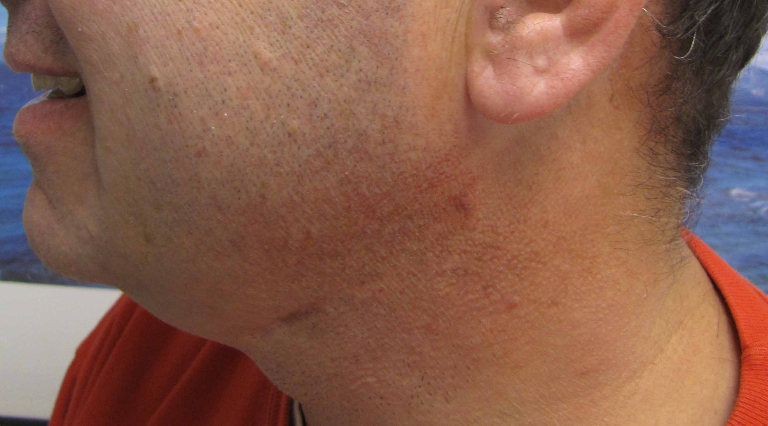
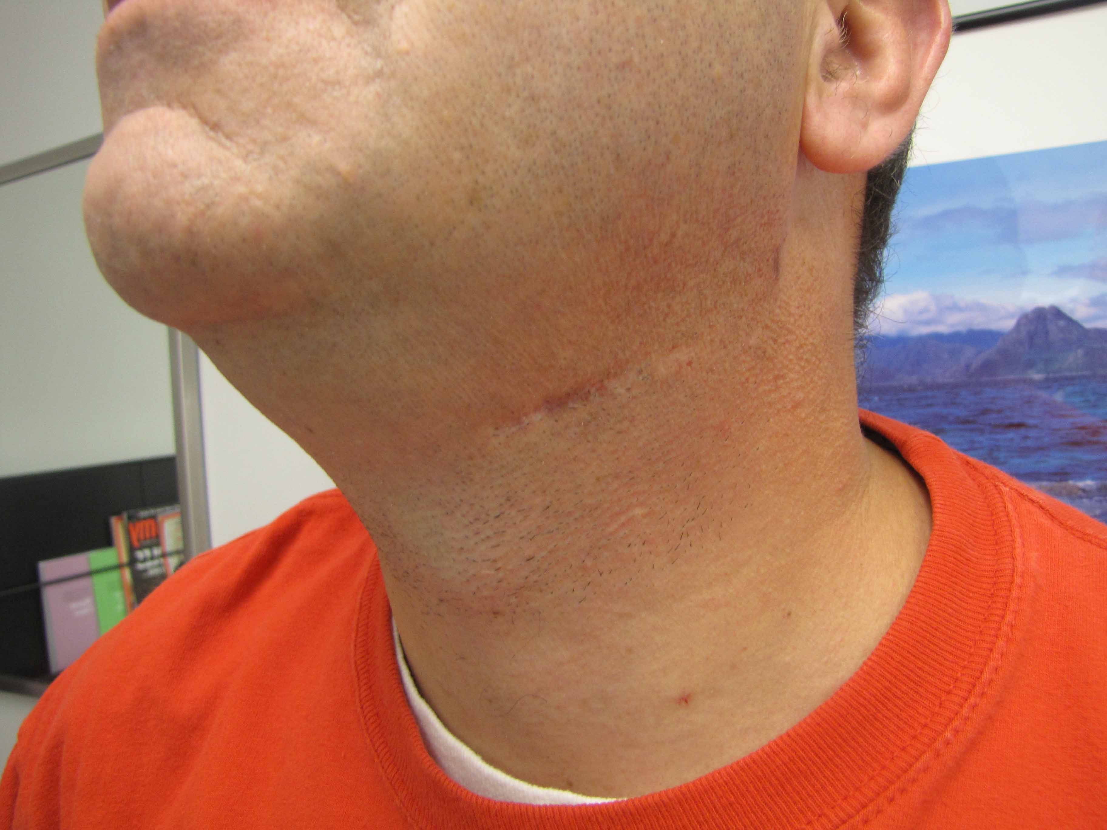
History: onset of left neck drainage beginning 2 1/2 months after left submandibular gland resection done for sialolithiasis
Modified Operative Note
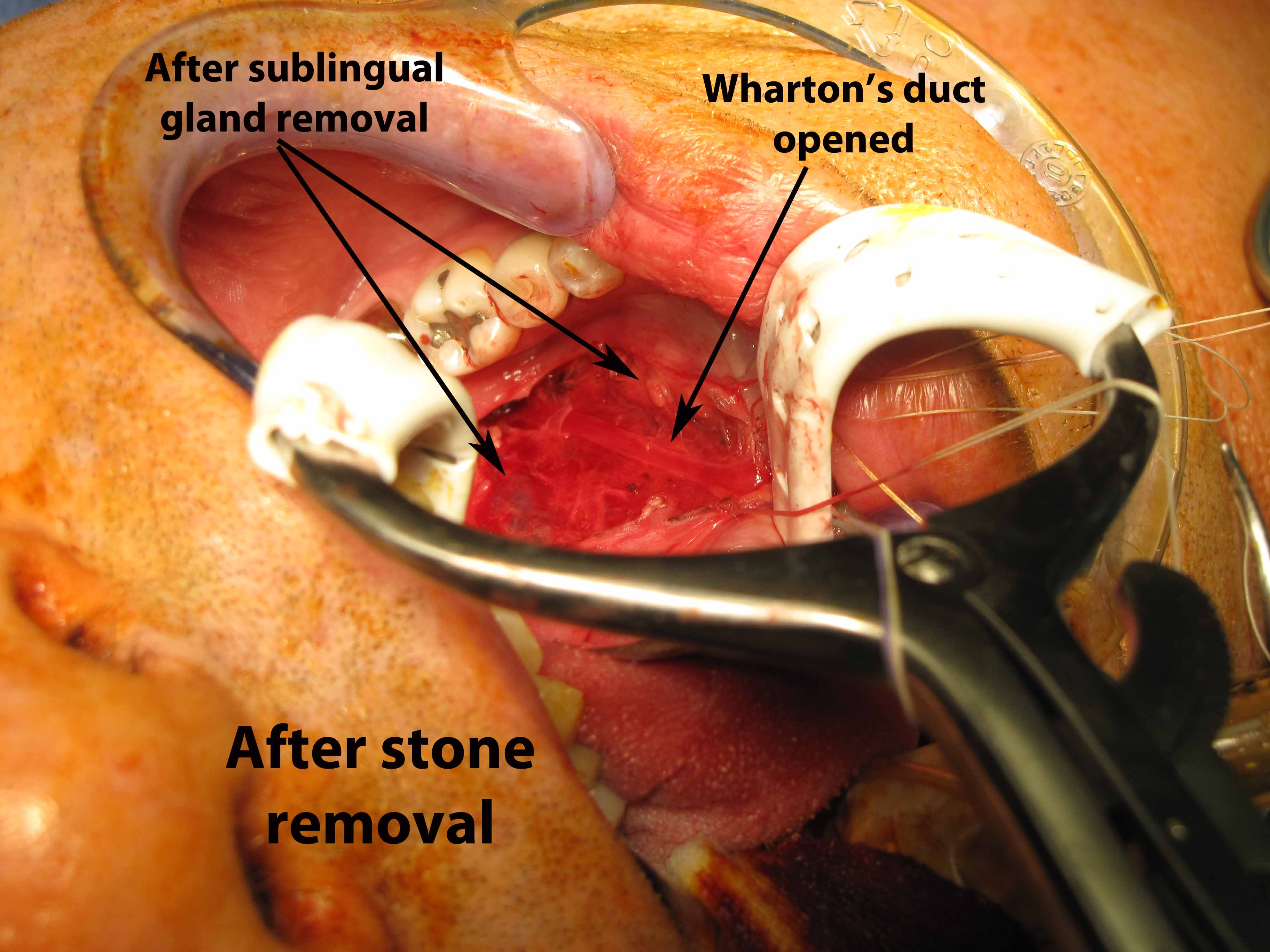
Preop Dx: Retained left ductal stone s/p left submandibular gland resection
Postop Dx: Same
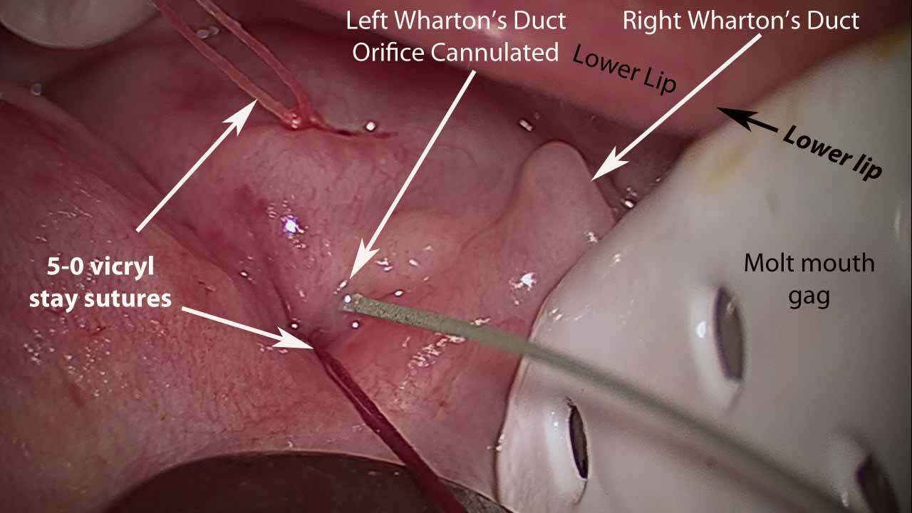
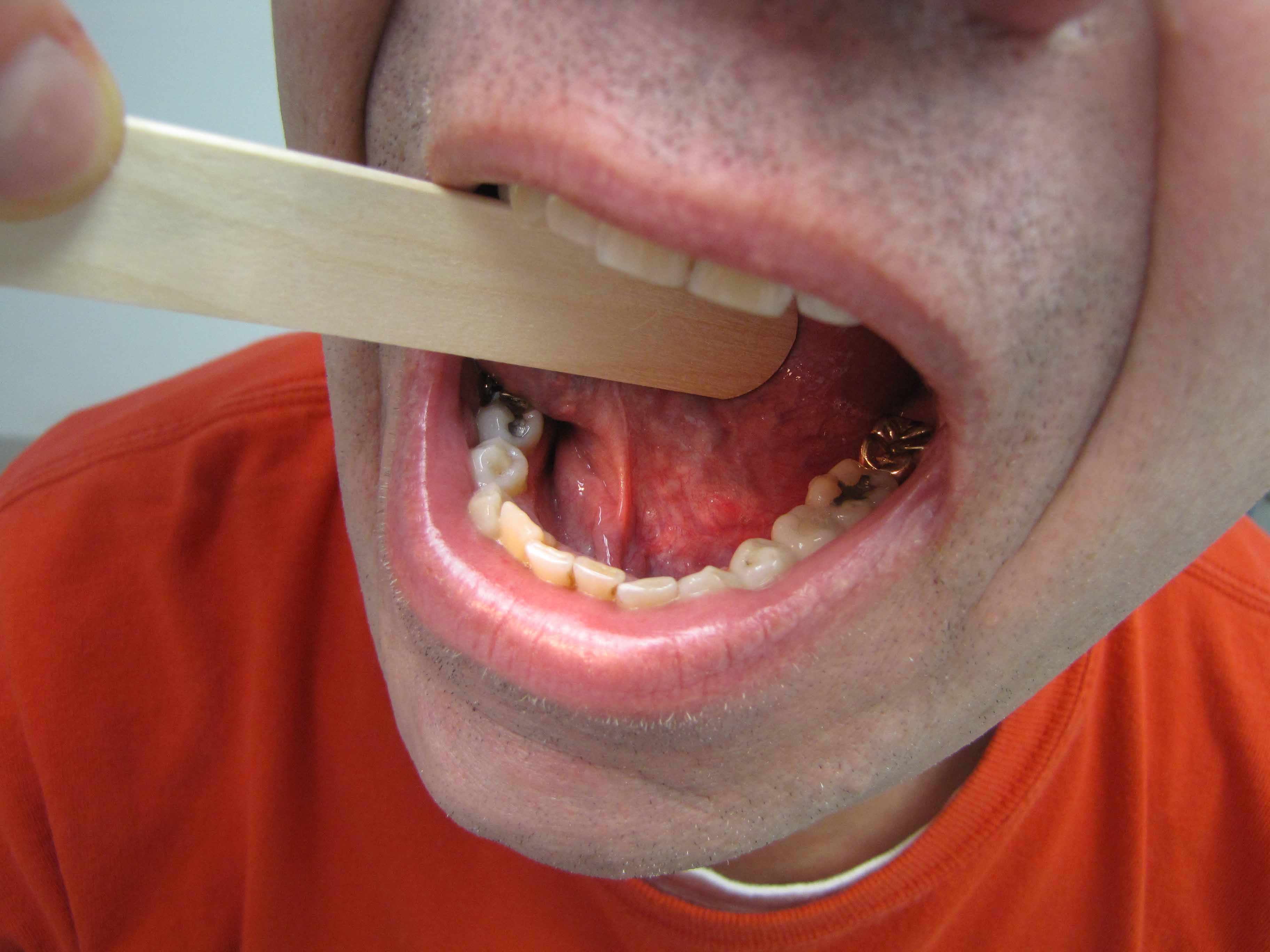
Procedure: Left submandibular sialodochoplasty (complex) with stone removal employing sialendoscopy
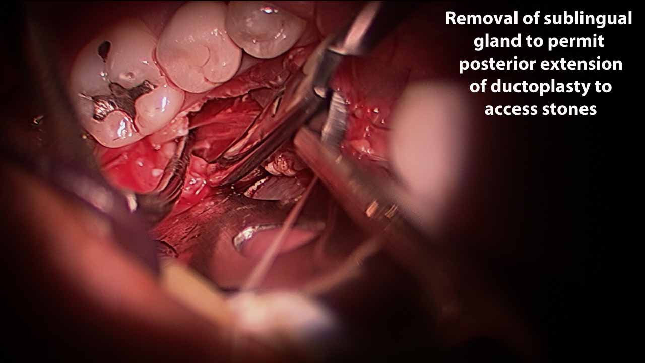
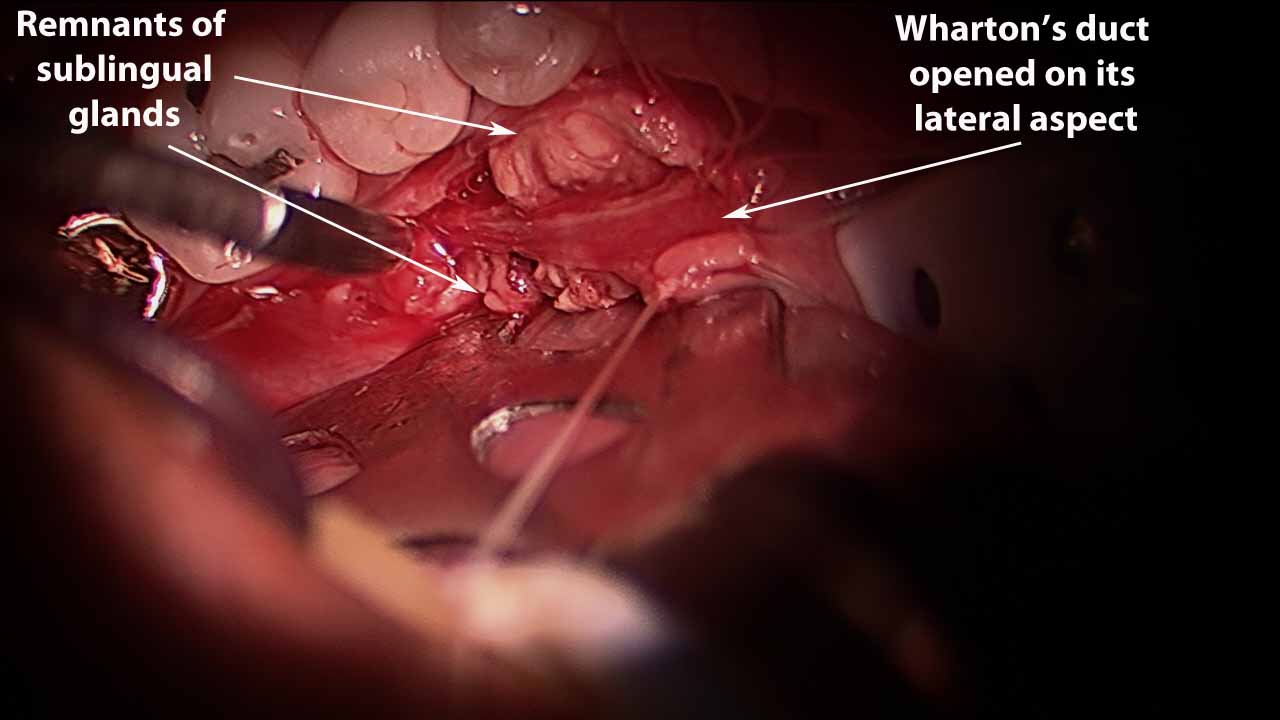
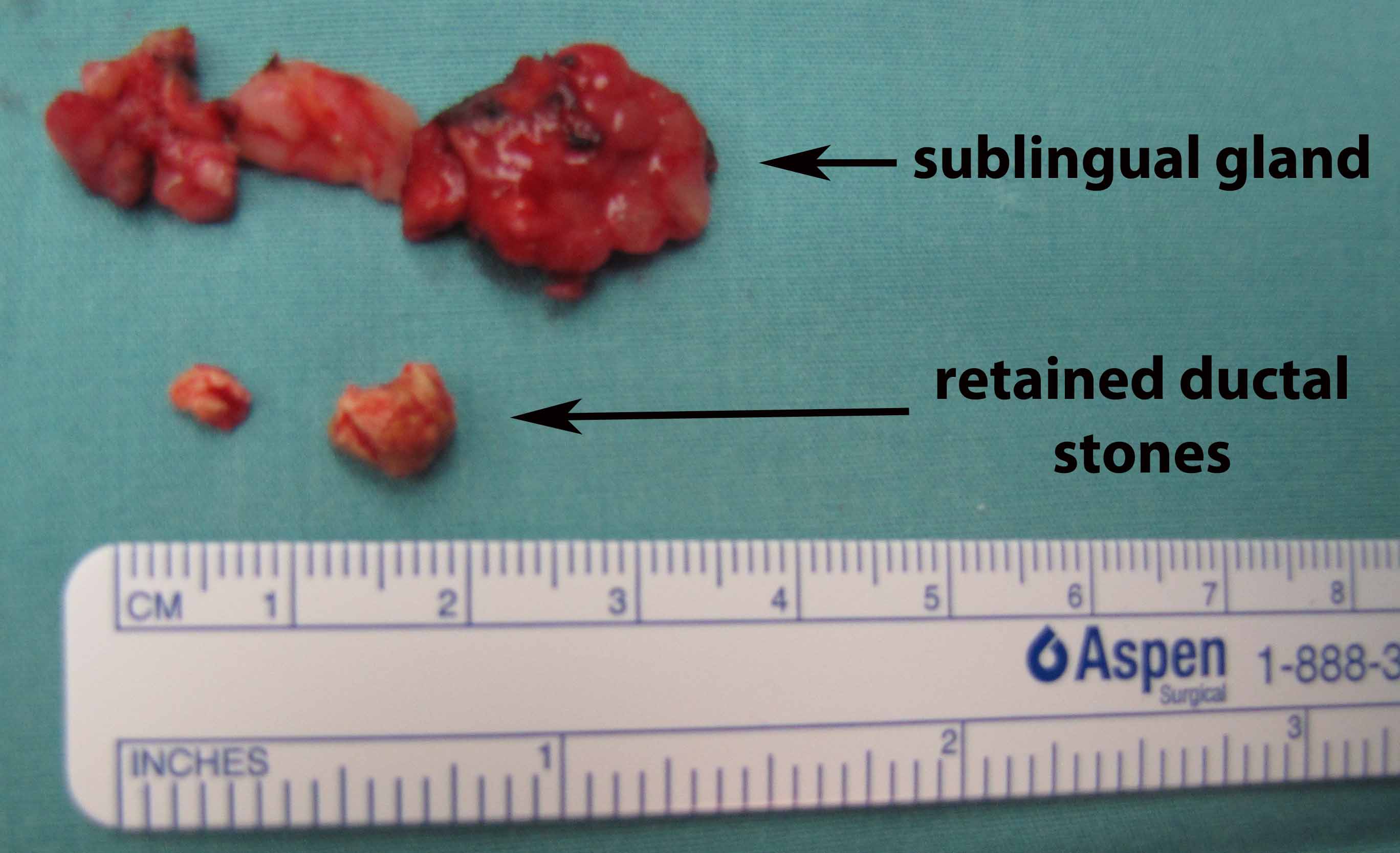
Left sublingual gland resection (subtotal)
Culture (tranoral) of submandibular site
Anesthesia: General OETT
Findings:
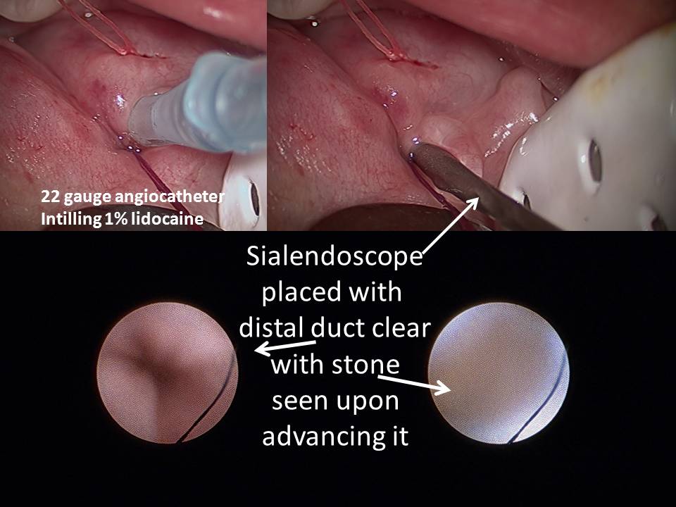
Duct readily canulated and then infused with 1% lidocaine
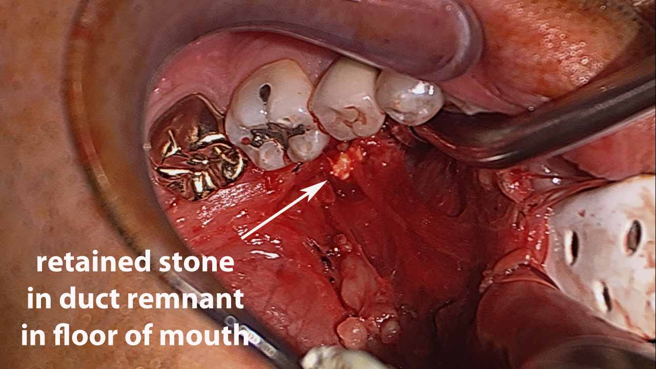
Serial dilation of duct orifice followed by sialendoscopy (diagnostic scope) to 4 cm from duct orifice where scarring of duct precluded clear view of stone (?stone vs scarring imaged)
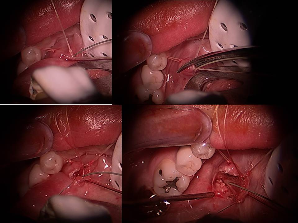
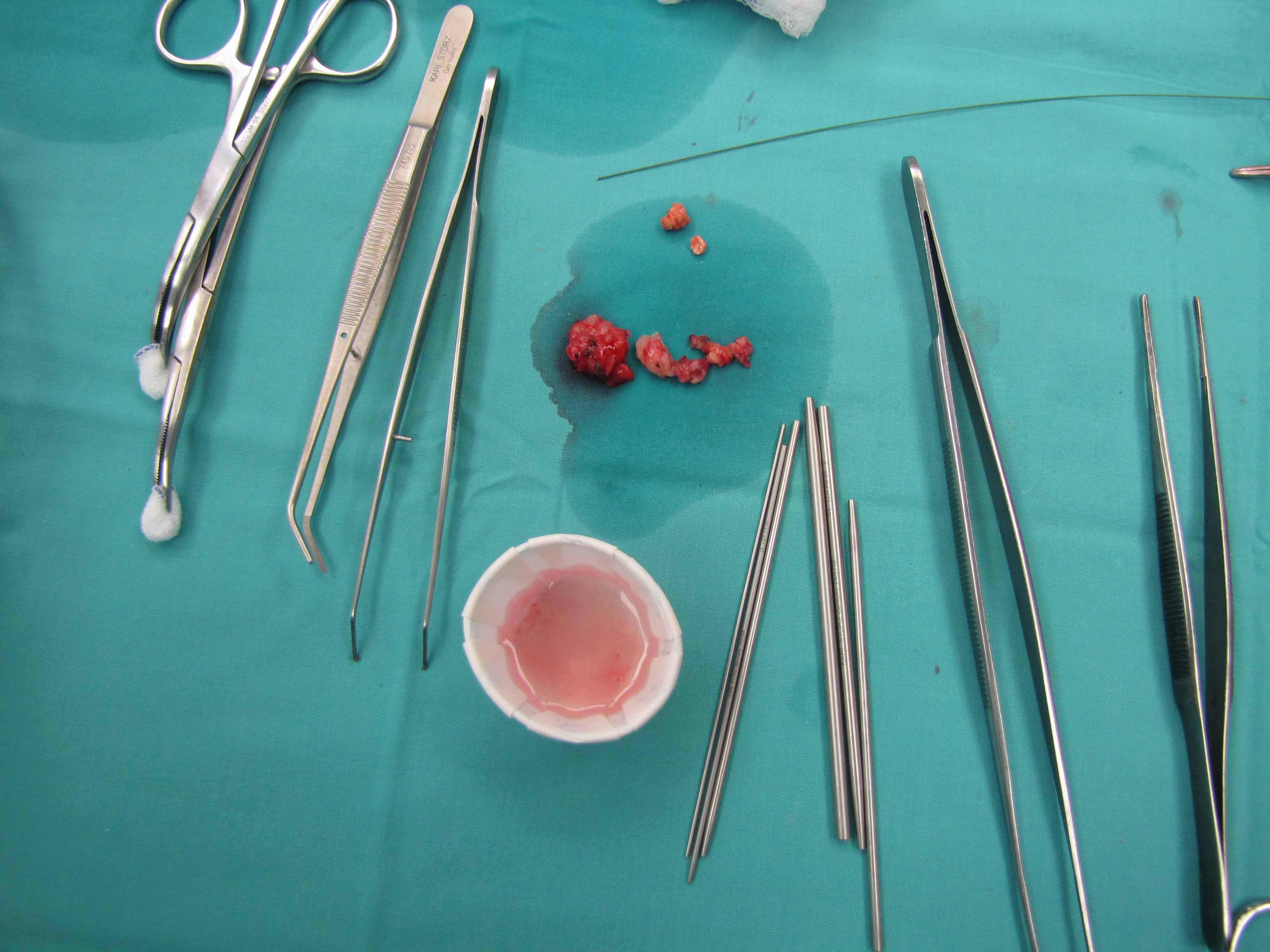
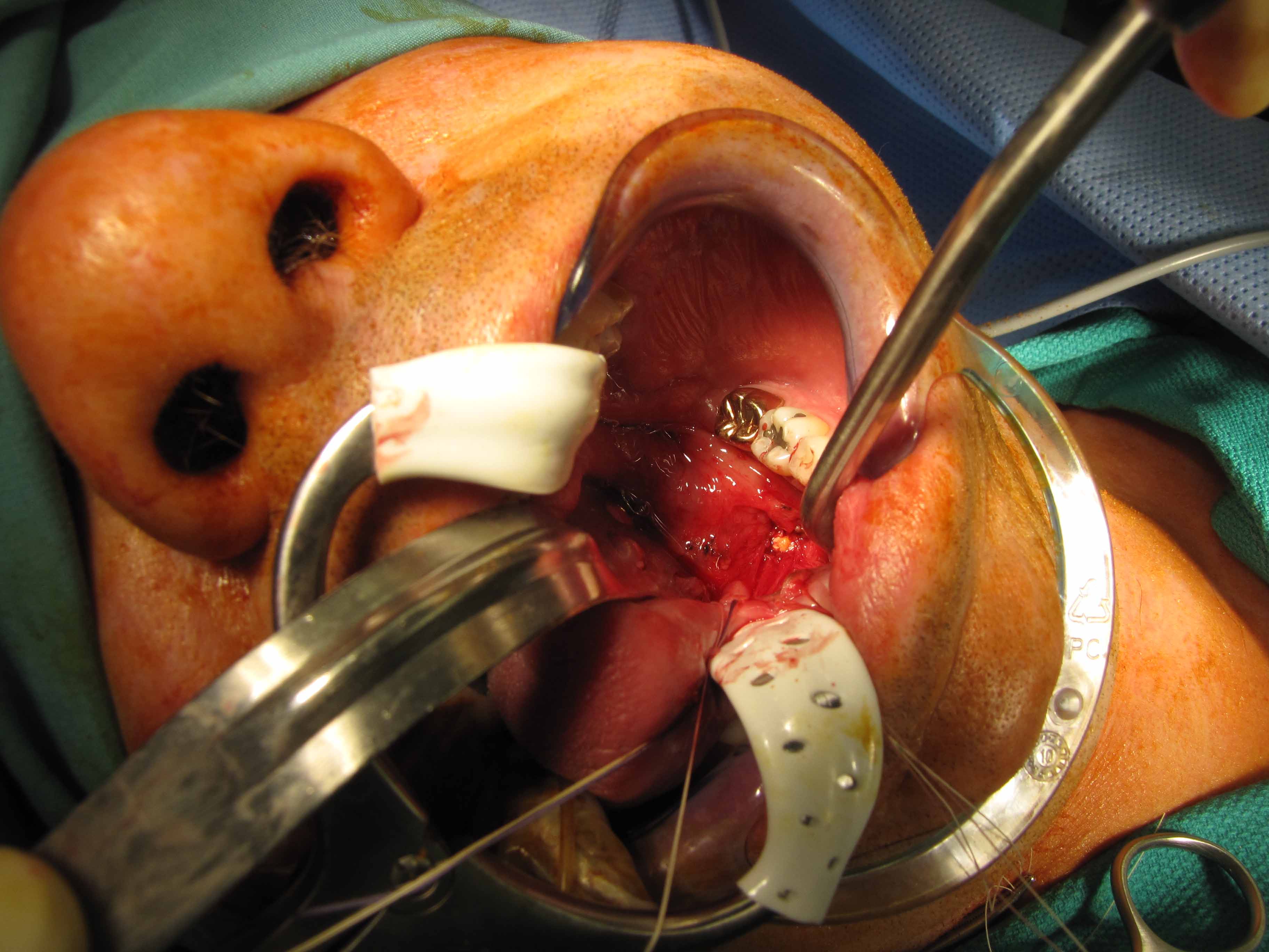
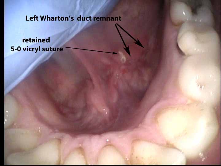
Duct opened with microscopic dissection with gradual removal of sublingual gland in course of marsupializing duct (sutured with 5-0 vicryl) -suturing done to ensure adequate intraoral drainage of site from which duct removed - in continuity with now opened duct with two large segments of stone removed. Copious irrigation - bipolar cautery followed by application of tannic acid. Specimens to path.