

click on image above to enlarge; advance with cursor over border
return to: Sialendoscopy; Sialendoscopy Tray
Salivary Gland Surgery Protocols; Sialolithiasis; Sialograms and Sialography; UCSF Sialendoscopy Nov 4 2015 Complex Cases
History
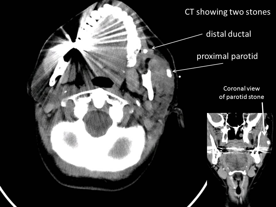
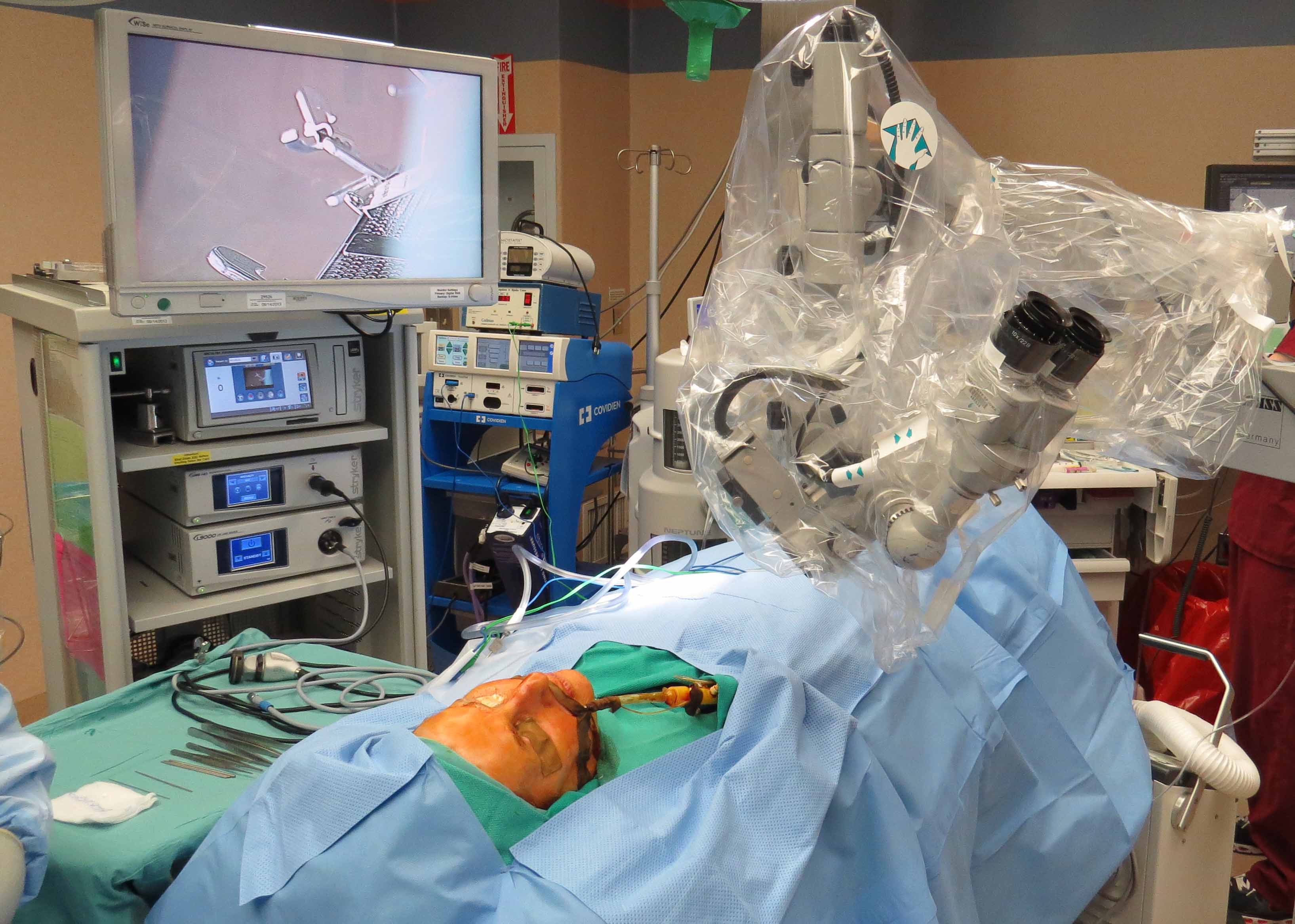
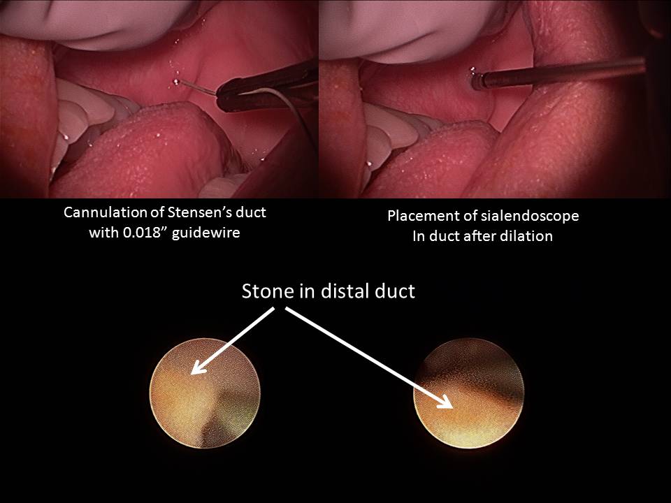
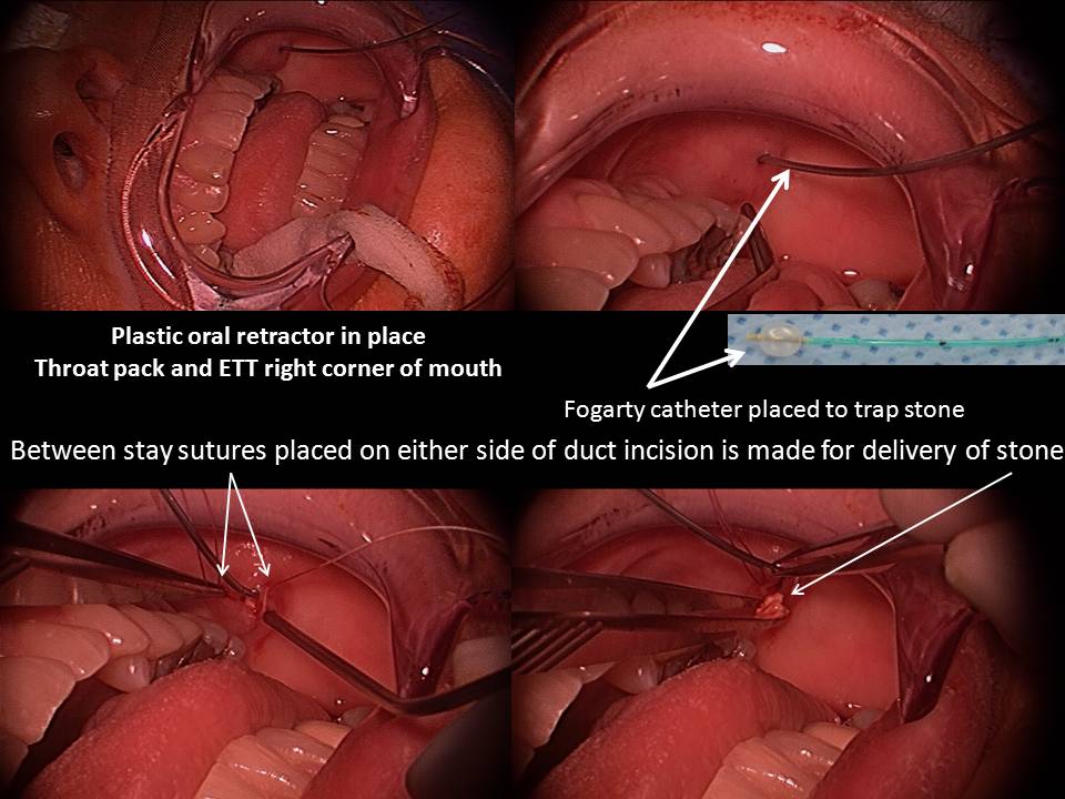
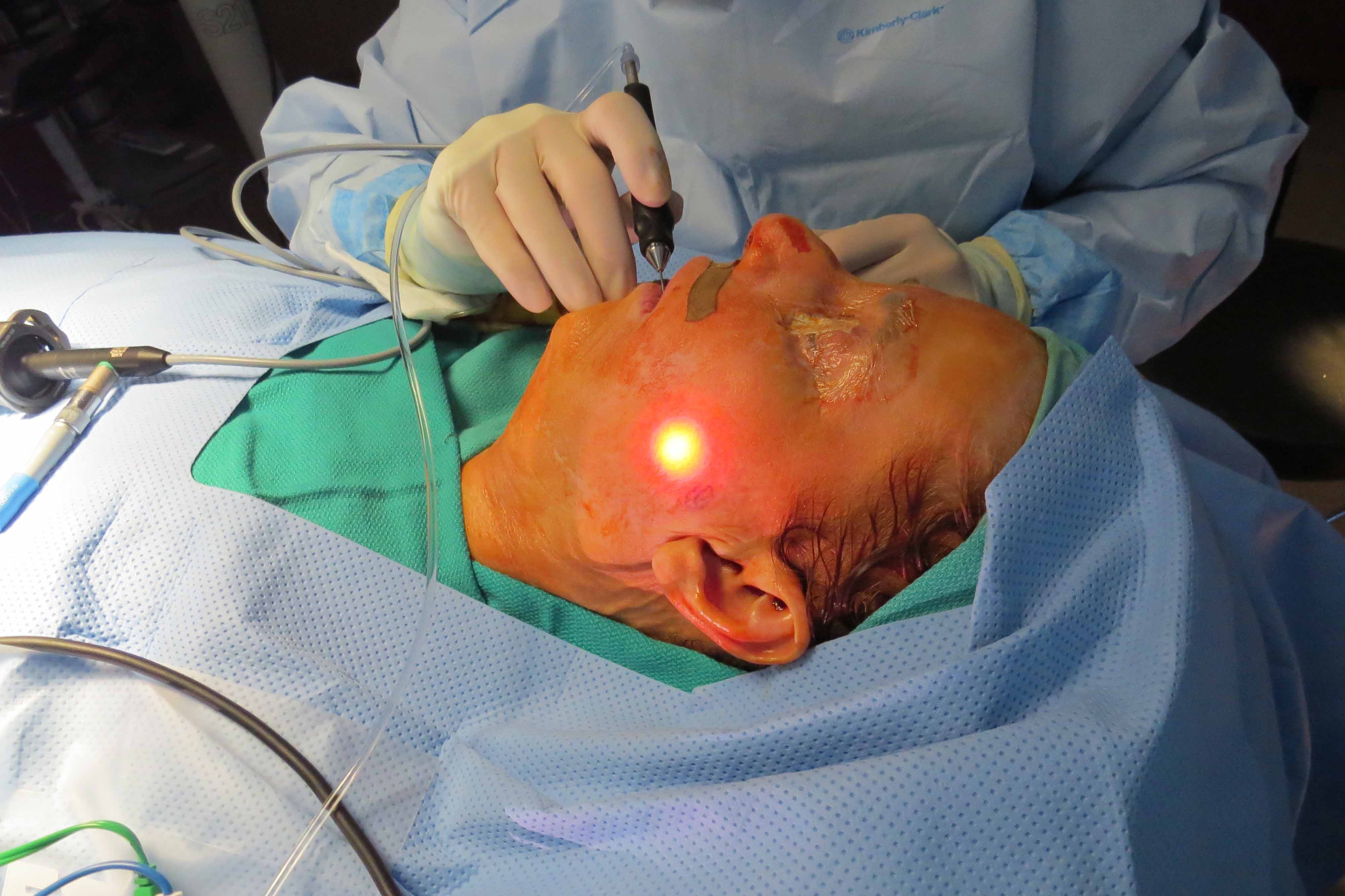
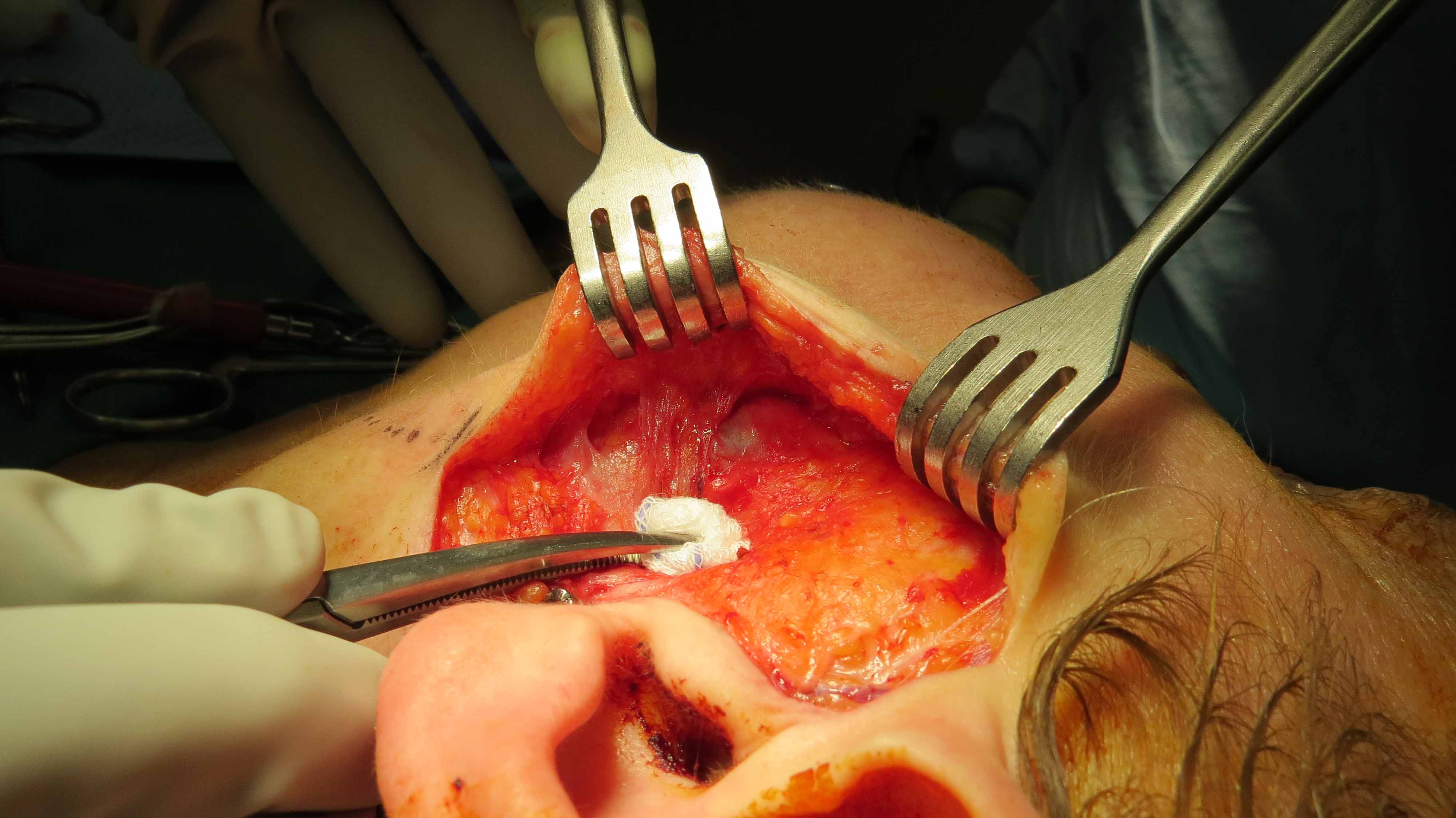
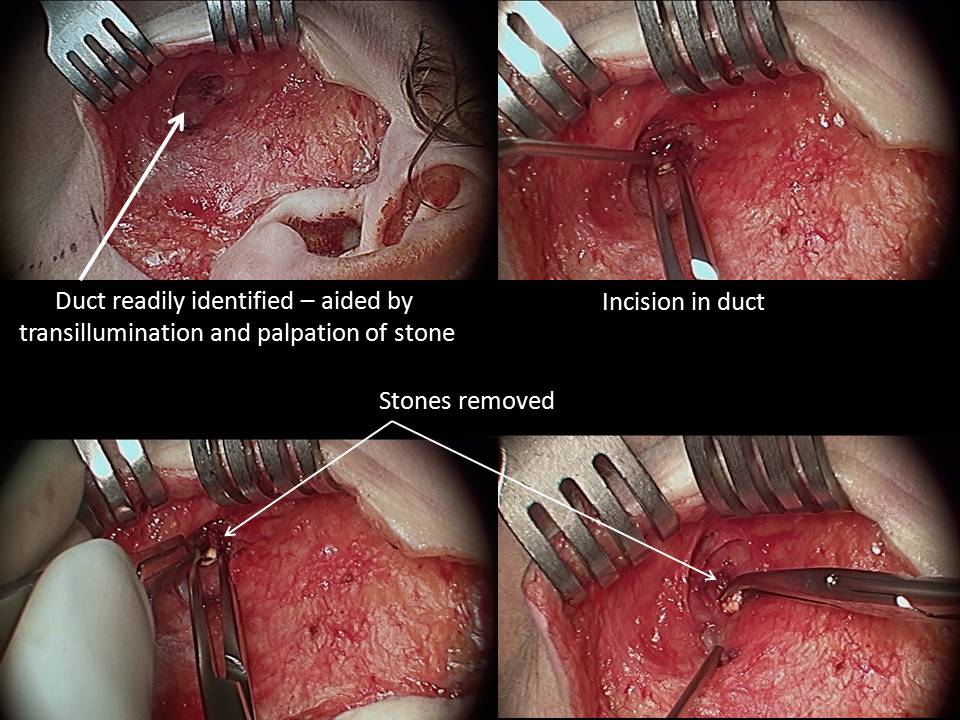
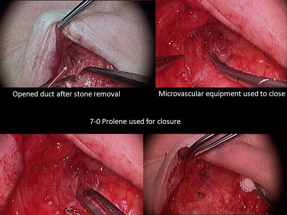
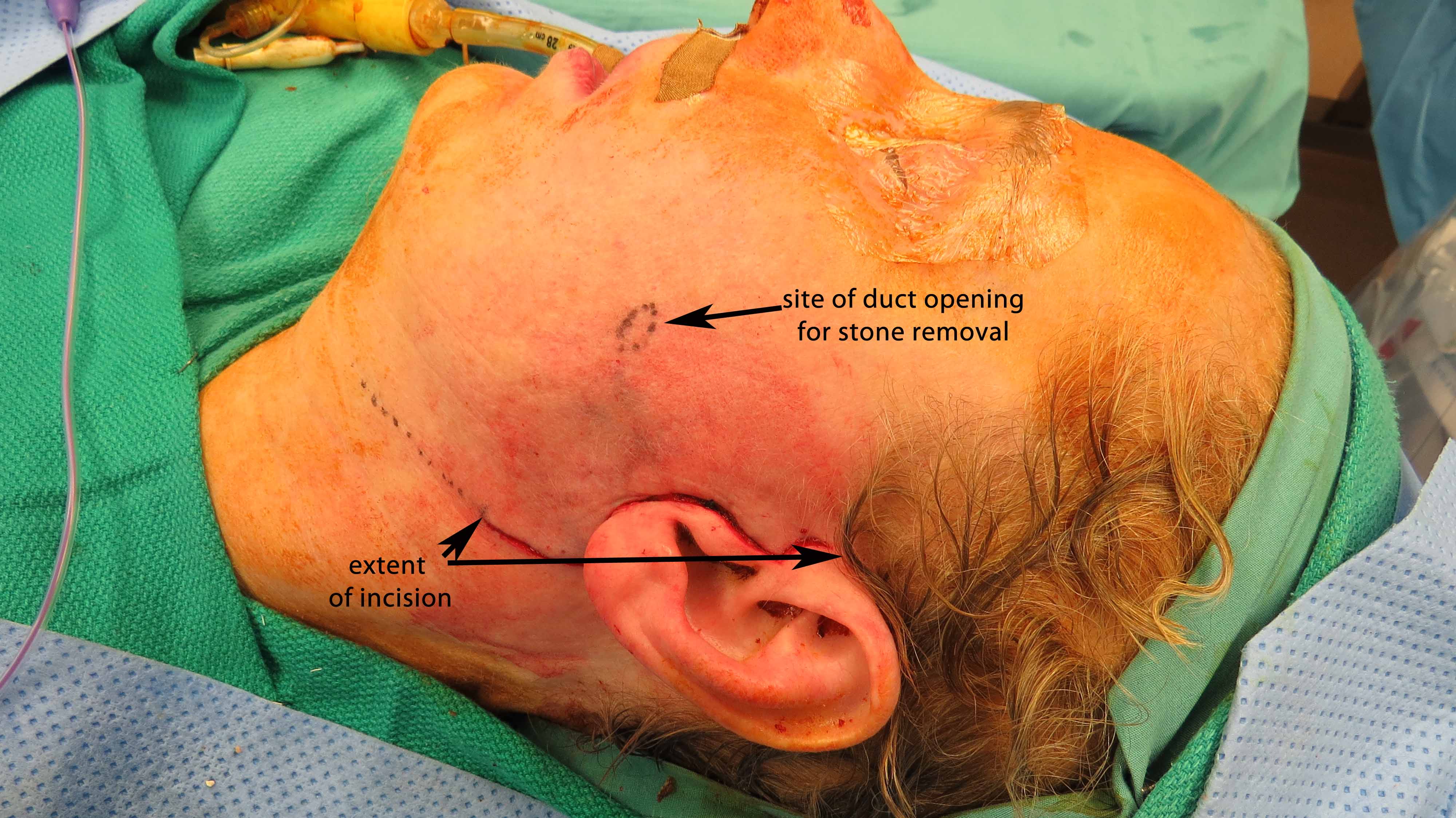
71 yo with left parotid swelling beginning 5 years previously requiring repeated courses of antibiotics. Two stones seen on CT are also palpable close to the hilum (palpable through cheek skin) and close to puncta intra-orally (palpable within the mouth). Salivary function preserving surgery was done with combined approach employing sialendoscopy as well as open approach to the duct via pre-auricular incision.
|
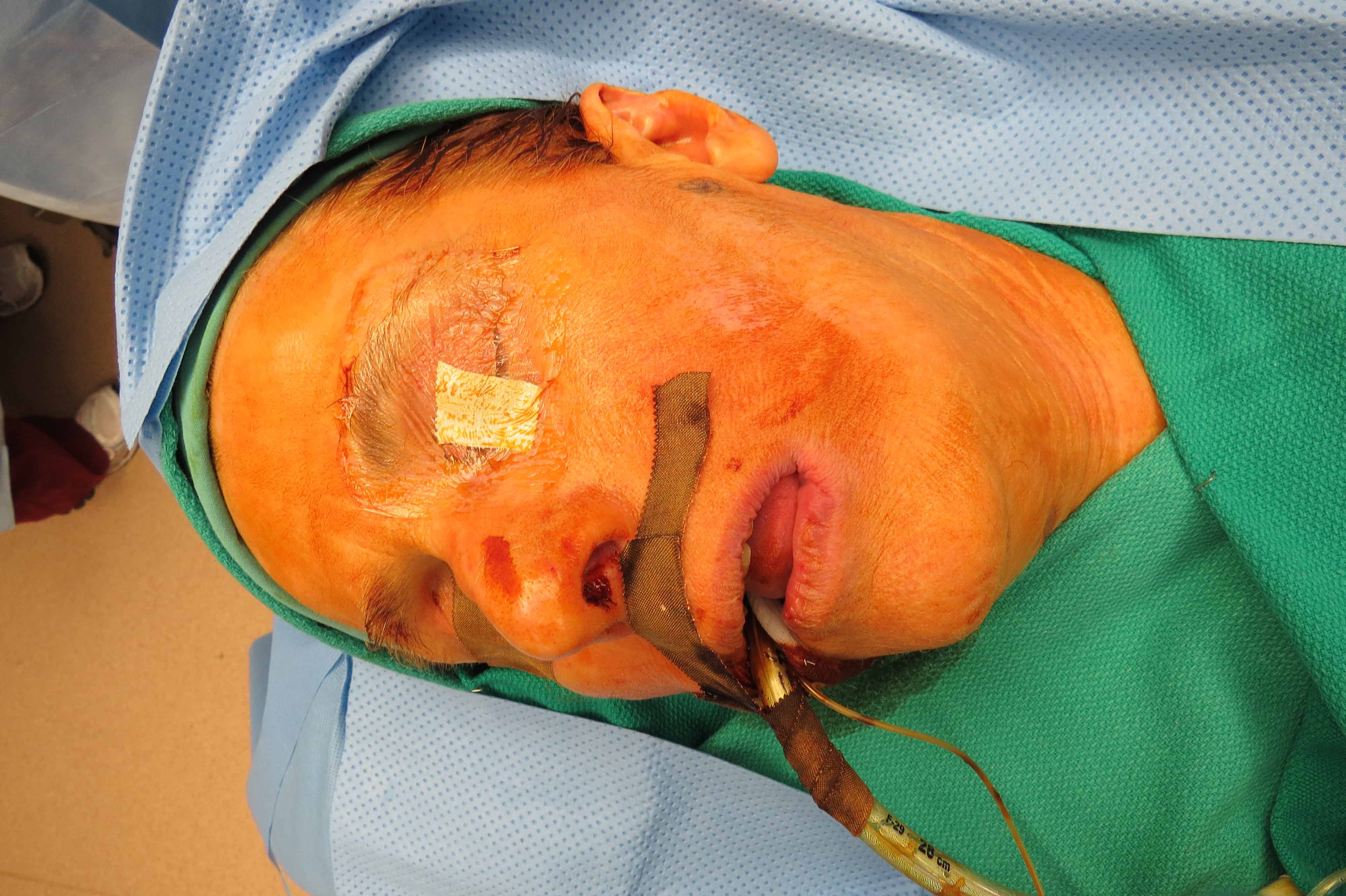
Followup after suture removal with good salivary flow from left Stensens duct |
flashmedia.uiowa.edu