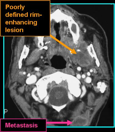
see also: Lipoma Radiology; Parotid Lipoma-like Liposarcoma (Salivary Swelling, Parotidectomy and Margin control re-excision with 10 year followup) - Clinical case example
- Malignant tumor derived from mesenchymal tissue with adipose differentiation (Barisella et al. 2020)
- Seen as fatty tumor with tissue stranding and nodularity
- Seen most commonly in the masticator space, larynx, and cervical neck
- Vast majority arise spontaneously, exceedingly few arise from lipoma
- Around 4% of all liposarcomas are found in the head and neck region (Scelsi et al. 2019)
- Seen more commonly in males and those between the fourth and seventh decade (Scelsi et al. 2019)
- Treatment involves wide surgical excision (Agarwal et al. 2017)
- On CT:
- Non-contrast: calcification seen in ~10% of cases
- Contrast: non-enhancing fatty lobules with septation if low grade, high grade enhances heterogenously and appears infiltrative
- On MR:
- T1 shows hyperintense fatty signal that appears dark with fat suppression
- T1 post-contrast shows heterogeneous enhancement of the cellular components, dark foci of necrosis
![]()

References
Barisella M, Giannini L, Piazza C. From head and neck lipoma to liposarcoma: a wide spectrum of differential diagnoses and their therapeutic implications. Curr Opin Otolaryngol Head Neck Surg. 2020 Apr;28(2):136-143. doi: 10.1097/MOO.0000000000000608. PMID: 32011399.
Agarwal J, Kadakia S, Agaimy A, Ogadzanov A, Khorsandi A, Chai RL. Pleomorphic liposarcoma of the head and neck: Presentation of two cases and literature review. Am J Otolaryngol. 2017 Jul-Aug;38(4):505-507. doi: 10.1016/j.amjoto.2017.04.012. Epub 2017 Apr 22. PMID: 28528729.
Scelsi CL, Wang A, Garvin CM, Bajaj M, Forseen SE, Gilbert BC. Head and Neck Sarcomas: A Review of Clinical and Imaging Findings Based on the 2013 World Health Organization Classification. AJR Am J Roentgenol. 2019 Mar;212(3):644-654. doi: 10.2214/AJR.18.19894. Epub 2018 Dec 27. PMID: 30589383.