click on image above to enlarge
return to: Otology - Neurotology
Note: Last updated before 2018
General Considerations for Bell’s palsy
- Signs and symptoms of Bell’s palsy
- Acute onset (<24-48 hours) of unilateral facial weakness or paralysis
- Loss of forehead wrinkling
- Brow ptosis
- Incomplete eyelid closure with possible exposure keratopathy
- Drooping of the mouth with possible drooling
- Absence of vesicles, dizziness, or hearing loss
- Acute onset (<24-48 hours) of unilateral facial weakness or paralysis
- Pathophysiology
- Neural edema and swelling within the Fallopian canal
- Likely related to Herpes Simplex Virus-1 (HSV-1) infection
- Neural compression and conduction blockade
- Labyrinthine segment of Fallopian (facial nerve) canal
- Particularly narrow (0.68 mm on average)
- Tight arachnoid band at opening of Fallopian canal at the fundus of the internal auditory canal
- Conduction blockade is located proximal to the geniculate ganglion (GG) in the vast majority of patients
- Labyrinthine segment of Fallopian (facial nerve) canal
- Initial management – see Bell’s Palsy Treatment Algorithm for tree diagram regarding the management of Bell’s palsy.
- Work-up
- Electrophysiologic testing
- Electroneurography (ENOG) if:
- Complete paralysis and 3-14 days from symptom onset
- Continue testing every 3 days until ENOG improves, or reaches 90% degeneration, or reaches 14 days
- Voluntary electromyography (EMG)
- Obtain if >90% degeneration on ENOG
- Voluntary motor unit potentials indicate good prognosis even if ENOG has >90% degeneration
- Electroneurography (ENOG) if:
- Imaging (CT or MRI) if:
- Clinical course, signs, and symptoms inconsistent with classic Bell’s palsy
- Gradual, progressive paralysis or waxing and waning course
- Presence of otorrhea, vestibular complaints, or hearing loss
- Severe paralysis for >6 months
- Serologic Lyme disease testing if:
- Vestibular symptoms, bilateral paralysis, or other suggestive signs or symptoms (e.g. rash)
- Electrophysiologic testing
- Conservative therapy
- Patients presenting <14 days after symptom onset
- Incomplete paralysis (paresis)
- Oral prednisone, 60-80 mg, once daily for 10-14 days
- Oral valcyclovir, 500 mg, 3 times daily for 10 days
- 5 day follow-up
- Complete paralysis
- If <3 days since onset treat with prednisone and valcyclovir, and follow-up on day 3 of complete paralysis with ENOG testing as mentioned above
- If 3-14 days since onset proceed with ENOG as above. Prednisone and acyclovir if <90% degeneration on ENOG
- Incomplete paralysis (paresis)
- Patients presenting >14 days after symptom onset with stable or improving motor function (including patients with complete paralysis)
- No therapy
- 6 month follow-up
- For all patients, ophthalmology consult if:
- Ocular symptoms are present
- Concern for decreased corneal sensation
- Prolonged paralysis expected
- Patients presenting <14 days after symptom onset
- Work-up
- Indications for surgical intervention
- Complete facial paralysis
- Presentation within 14 days of onset of complete paralysis
- >90% denervation on ENOG
- No voluntary EMG activity
- Patient fit and willing to undergo surgery
- Middle cranial fossa (MCF) approach
- Provides access to facial nerve from brainstem to the tympanic segment of Fallopian canal
- The only surgical approach with evidence supporting its efficacy for Bell’s palsy

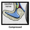
Figure 1. Schematic showing facial nerve decompression. Panel A shows a normal facial nerve within the Fallopian canal of the temporal bone. Panel B shows a swollen/edematous facial nerve compressed within the labyrinthine segment of the Fallopian canal. Panel C shows the facial nerve after decompression of the labyrinthine segment of the Fallopian canal. Figure reprinted with permission from Patricia Duffel, Eyerounds.org (Andresen 2017).
Preoperative Preparations – see General Considerations of Otologic Surgery for information regarding patient preparation, positioning, and facial nerve monitoring
- Imaging
- MRI with T1 post and CISS sequences
- BJG - Stenver’s view radiographs
- Orders to be placed preoperatively
- Cefazolin (Ancef ) and dexamethasone (Decadron) – given during induction
- Mannitol (0.5 mg/kg) – incision
- MRH - acyclovir – anytime
- Patient preparation and positioning as detailed elsewhere (link above)
- Facial nerve monitoring as described elsewhere (link above)
- Intraoperative ABR is not routinely used
Nursing Considerations
Anesthesia Considerations - see Otology Antibiotic Administration Guidelines for more detailed information regarding antibiotics
- Discuss with anesthesia team
- Cefazolin (Ancef) and dexamethasone (Decadron) – given during induction
- Mannitol (0.5 g/kg) – incision
- MRH - acyclovir – anytime
- Hyperventilate to end tidal CO2 of <30mmH2O until closing
- No paralytics
- Arterial line after patient is turned
Operative Procedure
- General principles of bony neural decompression
- Always drill parallel to the nerve
- Remove bone surrounding nerve with diamond burrs
- Once bone is egg shell thin, remove final layer of bone manually with hooks, Fisch nerve dissectors, etc.
- Incision and elevation of skin flaps
- BJG – postauricular posteriorly-based skin flap
- MRH - preauricular anteriorly-based skin flap
- Use 15 blade scalpel and incise to depth of temporoparietal (TP) fascia
- Elevate skin flap in plane of TP fascia
- Wrap skin flap in moist Raytech to prevent dessication during case
- Use tie-back suture to retract skin flap
- Beware of the frontal branch of the facial nerve
- Located within TP fascia proximally
- Deep to temporal fascia along zygomatic arch
- Fascia harvest
- Harvest 6x5 cm piece of temporalis fascia for later use
- Leave 1 cm rim of fascia along muscle edge to suture to during closure
- Incise the fascia sharply and dissect free of underlying muscle with scissors
- Harvest 6x5 cm piece of temporalis fascia for later use
- Muscle flap elevation
- Elevate the temporalis muscle off of the calvarium using a periosteal elevator
- Wrap the muscle in a moist Raytech to prevent dessication
- Retract with stay suture
- Elevate the temporalis muscle off of the calvarium using a periosteal elevator
- Creation of temporal craniotomy/bone flap
- 4x5 cm bone flap should be centered over the zygomatic root
- Zygomatic root estimates level of floor of middle fossa
- Make craniotomy with a cutting burrs
- Bone will be thinner anteroinferiorly and thicker posteriorly
- Once dura is seen below a thin layer of bone, switch to a diamond burr to avoid injury to the dura
- Remove the bone flap and set aside for later
- Freshen the edges of the craniotomy with the diamond burr
- Inner and outer cortical tables should be parallel to keep the House-Urban retractor from shifting/rocking
- May need to remove excess bone using ronguer or drill to access the middle fossa floor
- Especially true inferiorly
- 4x5 cm bone flap should be centered over the zygomatic root
- Retract temporal lobe and place House-Urban retractor
- Carefully dissect the dura off of the middle fossa floor in a posterior to anterior and lateral to medial direction
- Limits of exposure are the petrous ridge posteromedially and the foramen spinosum anteriorly
- Foramen spinosum is traversed by the middle meningeal artery
- Middle meningeal may need to be cauterized to provide adequate exposure
- Dissect dura off the greater superficial petrosal nerve (GSPN) carefully to avoid avulsion from the geniculate ganglion
- The geniculate ganglion (GG) may be dehiscent 5-15% of the time
- Limits of exposure are the petrous ridge posteromedially and the foramen spinosum anteriorly
- Dural reflections should be cauterized with bipolar forceps and then transected sharply
- House-Urban retractor is placed to retract the temporal lobe with the retractor blade tip carefully placed at the medial margin of the petrous ridge
- Carefully dissect the dura off of the middle fossa floor in a posterior to anterior and lateral to medial direction
- The superior aspect of the internal audiotory canal is identified as a line that bisects the GSPN and the arcuate eminence/ superior semicircular canal (SCC).
- Identify and ‘blue line’ the superior semicircular canal (SCC) by slowly drilling down the arcuate eminence
- Locating the yellow-white, dense bone of the otic capsule identifies the SCC
- The roof of the epitympanum is removed to expose the head of the malleus, cochleariform process and tympanic segment of the facial nerve.
- The bone over the GG is removed.
- Opening of the internal auditory canal (IAC) and Fallopian canal
- Skeletonize the IAC 180-270º at the porus
- All bone from the porous to Bill’s bar is removed. A rim of bone is left at porous to sustain the retractor blade
- Meatus of labyrinthine segment is identified at Bill’s bar and bony canal is removed from meatal foramen to GG
- Bone removal over labyrinthine segment is only 90º of canal
- Reduces risk of inadvertent injury to basal turn of cochlea and superior SCC ampulla
- Fallopian canal is then opened to the cochleariform process
- Thin layer of bone left over course of nerve until entire nerve is exposed
- Thin bony later is removed with small right angle hooks or Fisch nerve dissector
- A microscalpel (Beaver No. 59-10) is used to open the IAC from proximal to distal
- The tight arachnoid band at the meatal foramen is incised
- Intraoperative EEMG
- Performed with Prass probe or Parsons-McCabe facial stimulator
- Successively stimulate intrameatal through the tympanic segments
- Scrub nurse should visualize the forehead, eye, mouth, and chin to serve as a back-up to intra-operative EMG
- Closure
- Irrigate and ensure hemostasis intracranially
- IAC defect repair
- BJG – use temporalis fascia and temporalis muscle plug in petrous defect; no fat graft
- Temporalis muscle harvested from undersurface of temporalis muscle
- MRH – use abdominal fat (size of first thumb phalange) harvested from left inferior quadrant
- Use lateral Pfannensteil incision at bikini line
- Can later use fat sat imaging on surveillance MRI
- No penrose
- Close donor site with vicryl and monocryl sutures, as well as steristrips
- Apply pressure dressing with elastoplast and fluffs
- BJG – use temporalis fascia and temporalis muscle plug in petrous defect; no fat graft
- Resurface middle fossa floor with temporalis fascia.
- Place bone grafts over epitympanum and any other middle fossa floor defects. Bone grafts should be placed between fascial and dural layers.
- Place two dural elevation sutures inferiorly in the craniotomy window
- Replace bone flap
- BJG – no mini plates
- MRH - resorb-X plating system
- Plates placed on superior, posterior, and anterior part of bone flap
- Replace temporalis muscle
- Close subcutaneous tissue and skin
- Close the subcutaneous tissues with interrupted 3-0 vicryl sutures
- BJG - close skin with interrupted 3-0 nylon
- MRH - close skin with running/locking 3-0 nylon.
- Place bulky mastoid-type dressing with adequate pressure to help prevent subgaleal fluid from developing
Post-operative Care – see Postoperative Care Map for Skull Base Surgery for general information regarding post-operative care
- General post-operative care as per link above
- Bell’s palsy specific post-operative care
- Eye care
- Ophthalmology consult as indicated and mentioned above
- Artificial tears and lubricating ointment
- Temporary (adhesive) or permanent upper eyelid weight if indicated
- Temporary or permanent tarsorrhaphy if indicated
- Physical therapy
- Includes exercises, massage, acupuncture, and biofeedback
- Electrical stimulation generally NOT be used
- Eye care
Sample Dictation
The patient was brought to the operating room. General anesthesia was induced. A preinduction timeout was called and all present were in agreement. The patient was administered general anesthesia by the anesthesia team. Please see their note for full details. The right side of the head, which was marked, was then shaved in the standard fashion. A curvilinear preauricular incision was then marked in the standard fashion, infiltrated with 5 mL of 1% lidocaine with 1:100,000 epinephrine. The patient had facial nerve monitoring electrodes placed in the orbicularis oculi and orbicularis oris muscles, and these were found to be working properly with the appropriate impedances and tap stimulation. The patient was then turned 180 degrees clockwise away from anesthesia. An arterial line was placed. The right side of the head and abdomen were prepped and drapped.
A second timeout was then called and all present were in agreement, after discussing the side, site, procedure performed, allergies and antibiotics were all confirmed. The previously marked incision was incised with #15 blade, and dissection was carried down to the temporoparietal fascia. The skin flap was elevated anteriorly and the temporalis fascia graft was then harvested and placed aside in Ringer’s solution. The temporalis muscle was then incised and reflected inferiorly. A 5x5 cm temporal craniotomy centered on the zygomatic root was created. The bone flap was elevated without any evidence of dural tears. The dura was elevated from the patient's middle cranial fossa floor in a posterior-to-anterior direction. The greater superficial petrosal nerve was identified as well as the petrous ridge. The retractor was then placed along the petrous ridge at the center of the meatal plane. The anterior petrous apex was then drilled and found to be filled with bone marrow anterior to the IAC. The roof of the IAC was then skeletonized and the superior semicircular canal was identified and blue-lined. The tegmen tympani were then opened to expose the head of the malleus, the cochleariform process, and the tympanic segment of the facial nerve. Dissection was then carried in the IAC laterally until the geniculate ganglion and labyrinthine segment were completely identified, exposed and opened. Final egg-shelled bone was removed using 2.5 mm hooks and Fisch nerve dissectors. The band surrounding the meatal foramen was then lysed and the epineurium was then completely opened. The facial nerve was stimulated beginning in the intracanalicular portion and throughout the tympanic segment and found to have electrophysiologic parameters listed above. A fat graft was harvested from the patient's abdomen. This wound was then closed in layers and a Steri-Strip. Fat was then placed over the patient's dural defect in the IAC. The fascia graft was then placed over the cranial base. A bone chip was then fashioned from the craniotomy bone flap and placed over the tegmen defect. The dura was then suspended with a 4-0 nylon suture in 2 different locations. The bone flap was replaced and the Resorb plating system was used to affix the bone flap. The temporalis muscle was then replaced in its anatomic position and sutured in place in a watertight fashion. The galeal/deep dermal layer was closed in watertight fashion using 3-0 Polysorb sutures and the skin was then closed with a 3-0 running locking suture in continuous fashion. A mastoid dressing was then applied to the side of the head. All counts were correct at the end of the case. The patient was handed back over to anesthesia, where anesthetic was reversed. The patient was extubated and transferred to the surgical intensive care unit in stable condition.
References
Gantz B.J., Gmür A., Fisch U. Intraoperative evoked electromyography in Bell’s palsy. Am J Otolaryngol. 1982;3(4):273-278
Gantz BJ, Rubinstein JT, Gidley P, Woodworth GG. Surgical management of Bell's palsy. Laryngoscope 1999;109(8):1177-1188. https://www.ncbi.nlm.nih.gov/pubmed/10443817.
Welder JD, Allen RC, Shriver EM. Facial Nerve Palsy: Ocular Complications and Management. EyeRounds.org. posted July 14, 2015; Available from: http://www.EyeRounds.org/cases/215-facial-nerve.htm.
Facial Nerve Surgery, in Surgery of the Ear, M. Glasscock and G. Shambaugh, Editors. 2010, W.B. Saunders Company: Philadelphia.
Andresen NS, Clark TJE, Sun DQ, Hansen MR, Shriver EM. Bell's Palsy Treated With Facial Nerve Decompression, EyeRounds.org. posted August 1, 2017; Available from: http://EyeRounds.org/cases/256-Bells-Palsy.htm.
