return to: Sialograms and Sialography; Sialendoscopy; Salivary Gland Anatomic Anomalies and Foreign Bodies Lecture AHNS April 9 2013
see videos at bottom of page
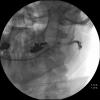
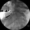
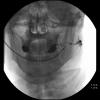
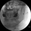
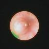
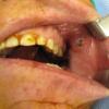
75 yo man relates that 2 years ago while running machinery he had a piece of metal enter into his left cheek at the corner of his mouth through the skin and had no problems from it at that time. A year later (one year ago) he (confirmed by his wife) related that he had an upper molar on the left infection that occurred coordinate with some swelling in his salivary gland. This episode was treated with antibiotics as well as a dental manipulation that allowed for both to resolve. Debate ensued about the etiology of the foreign body as either displaced dental amalgam (suggested by radiologist) versus slag from welding injury (supported by the patients wife as more consistent with timing). Further intermittent painful swelling of the left partoid gland warranted sialography followed by sialendoscopy with removal of the foreign body.
Procedure: Left parotid sialendoscopy with
- Removal of foreign body
- Complex ductoplasty with stent placement
- Kenalog infusion through stent into left parotid (3cc of kenalog 10)
Anesthesia: General
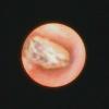
Findings: proximal duct narrowing addressed with progressive duct dilation over 0.018 inch guidewire - ultimately permitting removal of the ?slag/vs dental amalgam via 4-wire (0.4 mm) basket
Efforts before successful retrieval included:
- initial unsuccesful attempt with 4-wire (0.4 mm basket) without full dilation of proximal duct stenosis
- subsequent similar unsuccessful effort with 6 wire (0.6 mm basket) following full dilation of proximal stenosis (near puncta)
- fogarty catheter adjacent diagnostic scope (would not fit through large sialendoscope)
- two separate attempts at forceps to grab and pull through duct.
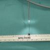
- final successful removal by trapping metallic foreign body in 4-wire (0.4 mm) basket and pulling through dilated duct orifice
8Fr pediatric feeding tube then placed over 0.18 inch guide -
- sutured in place
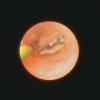
- then reassessed with intermediate scope showing it to be abutting first bifurcation
- therefore pulled back 1 cm more toward orifice and
- re-sutured with repeat sialendoscope through the stent showing good position.
- kenalog 10 then instilled through stent.
References
Blake Sullivan and Hoffman H: Dynamic imaging with sialography combined with sialendoscopy to manage a foreign body in Stensen's duct. Am J Otolaryngol. 2018 May - Jun;39(3):349-351. doi: 10.1016/j.amjoto.2018.03.001. Epub 2018 Mar 3
